 Movie
Movie Controller

[English] 日本語
 Yorodumi
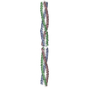
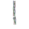
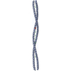
- PDB-6f5x: Crystal structure of the SYCP1 N-terminal head-to-head assembly i... -
+ Open data

- Basic information
| Entry | Database: PDB / ID: 6f5x | ||||||
|---|---|---|---|---|---|---|---|
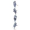
| Title | Crystal structure of the SYCP1 N-terminal head-to-head assembly in closed conformation | ||||||
 Components Components | Synaptonemal complex protein 1 | ||||||
 Keywords Keywords | STRUCTURAL PROTEIN / Meiosis / Chromosome structure / Coiled-coil / Self-assembly | ||||||
| Function / homology |  Function and homology information Function and homology informationtransverse filament / lateral element assembly / meiotic DNA repair synthesis / chiasma assembly / autosome / central element / sperm DNA condensation / synaptonemal complex assembly / homologous chromosome pairing at meiosis / synaptonemal complex ...transverse filament / lateral element assembly / meiotic DNA repair synthesis / chiasma assembly / autosome / central element / sperm DNA condensation / synaptonemal complex assembly / homologous chromosome pairing at meiosis / synaptonemal complex / reciprocal meiotic recombination / chromosome, centromeric region / Meiotic synapsis / male germ cell nucleus / regulation of protein localization / chromosome / spermatogenesis / double-stranded DNA binding / protein homotetramerization / cell division / DNA bindingSimilarity search - Function | ||||||
| Biological species |  Homo sapiens (human) Homo sapiens (human) | ||||||
| Method | X-RAY DIFFRACTION / SYNCHROTRON / SAD / Resolution: 1.91 Å | ||||||
 Authors Authors | Ratcliff, M. / Dunce, J.M. / Davies, O.R. | ||||||
| Funding support |  United Kingdom, 1items United Kingdom, 1items
| ||||||
 Citation Citation | Journal: Nat. Struct. Mol. Biol. / Year: 2018 Title: Structural basis of meiotic chromosome synapsis through SYCP1 self-assembly. Authors: Dunce, J.M. / Dunne, O.M. / Ratcliff, M. / Millan, C. / Madgwick, S. / Uson, I. / Davies, O.R. | ||||||
| History |
|
- Structure visualization
| Structure viewer | Molecule: MolmilJmol/JSmol |
|---|
- Downloads & links
-Download
| PDBx/mmCIF format | 6f5x.cif.gz | 48 KB | Display | PDBx/mmCIF format |
|---|---|---|---|---|
| PDB format | pdb6f5x.ent.gz | 33.9 KB | Display | PDB format |
| PDBx/mmJSON format | 6f5x.json.gz | Tree view | PDBx/mmJSON format | |
| Others |  Other downloads Other downloads |
-Validation report
| Arichive directory | https://data.pdbj.org/pub/pdb/validation_reports/f5/6f5xftp://data.pdbj.org/pub/pdb/validation_reports/f5/6f5x | HTTPS FTP |
|---|
-Related structure data


-Links
 PDBj
PDBj
- Assembly
| Deposited unit | 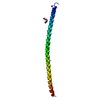
| |||||||||
|---|---|---|---|---|---|---|---|---|---|---|
| 1 | 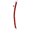
| |||||||||
| Unit cell |
| |||||||||
| Components on special symmetry positions |
|
-Components
| #1: Protein | / SCP-1 / Cancer/testis antigen 8 / CT8 Mass: 9168.571 Da / Num. of mol.: 1 Source method: isolated from a genetically manipulated source Source: (gene. exp.) Homo sapiens (human) / Gene: SYCP1, SCP1 / Production host:  Escherichia coli (E. coli) / References: UniProt: Q15431 Escherichia coli (E. coli) / References: UniProt: Q15431 | ||||
|---|---|---|---|---|---|
| #2: Chemical | Iodide  Mass: 126.904 Da / Num. of mol.: 2 / Source method: obtained synthetically / Formula: I Mass: 126.904 Da / Num. of mol.: 2 / Source method: obtained synthetically / Formula: I#3: Chemical | ChemComp-PGE / | Polyethylene glycol  Mass: 150.173 Da / Num. of mol.: 1 / Source method: obtained synthetically / Formula: C6H14O4 Mass: 150.173 Da / Num. of mol.: 1 / Source method: obtained synthetically / Formula: C6H14O4#4: Water | ChemComp-HOH / | Water Mass: 18.015 Da / Num. of mol.: 32 / Source method: isolated from a natural source / Formula: H2O Mass: 18.015 Da / Num. of mol.: 32 / Source method: isolated from a natural source / Formula: H2O |
-Experimental details
-Experiment
| Experiment | Method: X-RAY DIFFRACTION / Number of used crystals: 1 |
|---|
- Sample preparation
| Crystal | Density Matthews: 2.55 Å3/Da / Density % sol: 51.75 % |
|---|---|
| Crystal grow | Temperature: 293 K / Method: vapor diffusion, hanging drop / pH: 6.2 Details: 140 mM NaCl, 70 mM Na/K phosphate pH 6.2, 35% (v/v) PEG200; soaked in 40% (v/v) PEG200 and 100 mM NaI |
-Data collection
| Diffraction | Mean temperature: 100 K |
|---|---|
| Diffraction source | Source: SYNCHROTRON / Site: Diamond / Beamline: I02 / Wavelength: 1.7712 Å |
| Detector | Type: DECTRIS PILATUS 6M-F / Detector: PIXEL / Date: May 23, 2015 |
| Radiation | Protocol: SINGLE WAVELENGTH / Monochromatic (M) / Laue (L): M / Scattering type: x-ray |
| Radiation wavelength | Wavelength: 1.7712 Å / Relative weight: 1 |
| Reflection | Resolution: 1.91→41.44 Å / Num. obs: 7678 / % possible obs: 99.3 % / Redundancy: 5.9 % / Biso Wilson estimate: 33.34 Å2 / CC1/2: 1 / Rmerge(I) obs: 0.028 / Rpim(I) all: 0.017 / Rrim(I) all: 0.036 / Net I/σ(I): 27.9 |
| Reflection shell | Resolution: 1.91→1.95 Å / Redundancy: 3.7 % / Rmerge(I) obs: 0.678 / Mean I/σ(I) obs: 1.8 / Num. unique obs: 464 / CC1/2: 0.839 / Rpim(I) all: 0.541 / Rrim(I) all: 0.873 / % possible all: 92.1 |
- Processing
| Software |
| |||||||||||||||||||||||||||||||||||||||||||||||||
|---|---|---|---|---|---|---|---|---|---|---|---|---|---|---|---|---|---|---|---|---|---|---|---|---|---|---|---|---|---|---|---|---|---|---|---|---|---|---|---|---|---|---|---|---|---|---|---|---|---|---|
| Refinement | Method to determine structure: SAD / Resolution: 1.91→41.44 Å / SU ML: 0.27 / Cross valid method: FREE R-VALUE / σ(F): 2.02 / Phase error: 25.34 Details: Refined against data corrected for anisotropy (FP_ISOB/SIGFP_ISOB) using the UCLA diffraction anisotropy server.
| |||||||||||||||||||||||||||||||||||||||||||||||||
| Solvent computation | Shrinkage radii: 0.7 Å / VDW probe radii: 1 Å | |||||||||||||||||||||||||||||||||||||||||||||||||
| Displacement parameters | Biso mean: 58.4 Å2 | |||||||||||||||||||||||||||||||||||||||||||||||||
| Refinement step | Cycle: LAST / Resolution: 1.91→41.44 Å
| |||||||||||||||||||||||||||||||||||||||||||||||||
| Refine LS restraints |
| |||||||||||||||||||||||||||||||||||||||||||||||||
| LS refinement shell |
|
