 Movie
Movie Controller


+ Open data

- Basic information
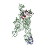
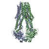
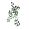
| Entry | Database: PDB / ID: 6f4a | ||||||
|---|---|---|---|---|---|---|---|
| Title | Yeast mitochondrial RNA degradosome complex mtEXO | ||||||
 Components Components |
| ||||||
 Keywords Keywords |  HYDROLASE / RNA degradation / mitochondria / nuclease / helicase / protein complex HYDROLASE / RNA degradation / mitochondria / nuclease / helicase / protein complex | ||||||
| Function / homology |  Function and homology information Function and homology informationmitochondrial chromosome / mitochondrial degradosome / mitochondrial RNA catabolic process / exoribonuclease II activity / mitochondrial DNA replication / Group I intron splicing / RNA nuclease activity / RNA helicase activity / RNA helicase / mitochondrion ...mitochondrial chromosome / mitochondrial degradosome / mitochondrial RNA catabolic process / exoribonuclease II activity / mitochondrial DNA replication / Group I intron splicing / RNA nuclease activity / RNA helicase activity / RNA helicase / mitochondrion / RNA binding / ATP bindingSimilarity search - Function | ||||||
| Biological species |  Candida glabrata (fungus) Candida glabrata (fungus) Escherichia coli (E. coli) Escherichia coli (E. coli) | ||||||
| Method | X-RAY DIFFRACTION / SYNCHROTRON / MOLECULAR REPLACEMENT / Resolution: 3.55 Å | ||||||
 Authors Authors | Razew, M. / Nowak, E. / Nowotny, M. | ||||||
| Funding support |  Poland, 1items Poland, 1items
| ||||||
 Citation Citation | Journal: Nat Commun / Year: 2018 Title: Structural analysis of mtEXO mitochondrial RNA degradosome reveals tight coupling of nuclease and helicase components. Authors: Razew, M. / Warkocki, Z. / Taube, M. / Kolondra, A. / Czarnocki-Cieciura, M. / Nowak, E. / Labedzka-Dmoch, K. / Kawinska, A. / Piatkowski, J. / Golik, P. / Kozak, M. / Dziembowski, A. / Nowotny, M. | ||||||
| History |
|
- Structure visualization
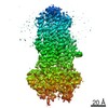
| Structure viewer | Molecule: MolmilJmol/JSmol |
|---|
- Downloads & links
-Download
| PDBx/mmCIF format | 6f4a.cif.gz | 434.4 KB | Display | PDBx/mmCIF format |
|---|---|---|---|---|
| PDB format | pdb6f4a.ent.gz | 338.1 KB | Display | PDB format |
| PDBx/mmJSON format | 6f4a.json.gz | Tree view | PDBx/mmJSON format | |
| Others |  Other downloads Other downloads |
-Validation report
| Arichive directory | https://data.pdbj.org/pub/pdb/validation_reports/f4/6f4aftp://data.pdbj.org/pub/pdb/validation_reports/f4/6f4a | HTTPS FTP |
|---|
-Related structure data
| Related structure data |  6f3hSC 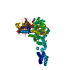 3rc3S S: Starting model for refinement C: citing same article ( |
|---|---|
| Similar structure data |


-Links
 PDBj
PDBj- Assembly
| Deposited unit | 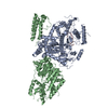
| ||||||||
|---|---|---|---|---|---|---|---|---|---|
| 1 |
| ||||||||
| Unit cell |
|
-Components
| #1: Protein | Mass: 94687.203 Da / Num. of mol.: 1 Source method: isolated from a genetically manipulated source Details: 69 aminoacid N-terminal truncation was introduced in the full length protein Source: (gene. exp.) Candida glabrata (fungus) / Gene: AO440_004157 / Production host: Escherichia coli (E. coli) / Variant (production host): RIL / References: UniProt: A0A0W0CXR7, UniProt: Q6FJE0*PLUS |
|---|---|
| #2: Protein | Mass: 73477.531 Da / Num. of mol.: 1 Source method: isolated from a genetically manipulated source Details: 42 aminoacid N-terminal truncation and 14 aminoacid C-terminal truncation was introduced to the full length protein Source: (gene. exp.) Candida glabrata (fungus) / Gene: SUV3, CAGL0L12386g / Production host: Escherichia coli (E. coli) / Variant (production host): RIL / References: UniProt: Q6FKD7 |
| #3: RNA chain | Mass: 1899.213 Da / Num. of mol.: 1 / Source method: isolated from a natural source / Details: RNA molecule co-purified with the protein / Source: (natural) Escherichia coli (E. coli) / Variant: RIL |
-Experimental details
-Experiment
| Experiment | Method: X-RAY DIFFRACTION / Number of used crystals: 1 |
|---|
- Sample preparation
| Crystal | Density Matthews: 3.3 Å3/Da / Density % sol: 62.76 % |
|---|---|
| Crystal grow | Temperature: 291 K / Method: vapor diffusion, hanging drop Details: 0.2M ammonium citrate tribasic (pH 7.0), 18% (w/v) PEG3350 |
-Data collection
| Diffraction | Mean temperature: 100 K |
|---|---|
| Diffraction source | Source: SYNCHROTRON / Site: ESRF  / Beamline: ID29 / Wavelength: 0.97625 Å / Beamline: ID29 / Wavelength: 0.97625 Å |
| Detector | Type: DECTRIS PILATUS 6M / Detector: PIXEL / Date: May 8, 2015 |
| Radiation | Protocol: SINGLE WAVELENGTH / Monochromatic (M) / Laue (L): M / Scattering type: x-ray |
| Radiation wavelength | Wavelength: 0.97625 Å / Relative weight: 1 |
| Reflection | Resolution: 3.55→50.1 Å / Num. obs: 26391 / % possible obs: 95.4 % / Redundancy: 4.3 % / Rrim(I) all: 0.033 / Net I/σ(I): 27.4 |
| Reflection shell | Resolution: 3.55→3.64 Å / Rrim(I) all: 0.225 |
- Processing
| Software |
| ||||||||||||||||||||||||||||||||||||||||||||||||||||||||||||||||||||||
|---|---|---|---|---|---|---|---|---|---|---|---|---|---|---|---|---|---|---|---|---|---|---|---|---|---|---|---|---|---|---|---|---|---|---|---|---|---|---|---|---|---|---|---|---|---|---|---|---|---|---|---|---|---|---|---|---|---|---|---|---|---|---|---|---|---|---|---|---|---|---|---|
| Refinement | Method to determine structure: MOLECULAR REPLACEMENT Starting model: 6F3H, 3RC3 Resolution: 3.55→49.061 Å / SU ML: 0.53 / Cross valid method: FREE R-VALUE / σ(F): 1.38 / Phase error: 37.64 / Stereochemistry target values: MLHL
| ||||||||||||||||||||||||||||||||||||||||||||||||||||||||||||||||||||||
| Solvent computation | Shrinkage radii: 0.9 Å / VDW probe radii: 1.11 Å / Solvent model: FLAT BULK SOLVENT MODEL | ||||||||||||||||||||||||||||||||||||||||||||||||||||||||||||||||||||||
| Refinement step | Cycle: LAST / Resolution: 3.55→49.061 Å
| ||||||||||||||||||||||||||||||||||||||||||||||||||||||||||||||||||||||
| Refine LS restraints |
| ||||||||||||||||||||||||||||||||||||||||||||||||||||||||||||||||||||||
| LS refinement shell |
| ||||||||||||||||||||||||||||||||||||||||||||||||||||||||||||||||||||||
| Refinement TLS params. | Method: refined / Origin x: -3.4509 Å / Origin y: -51.2107 Å / Origin z: 31.8815 Å
| ||||||||||||||||||||||||||||||||||||||||||||||||||||||||||||||||||||||
| Refinement TLS group | Selection details: all |
