 Movie
Movie Controller

[English] 日本語
 Yorodumi
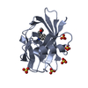
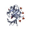
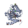
- PDB-6f20: Complex between MTH1 and compound 1 (a 7-azaindole-4-ester derivative) -
+ Open data

- Basic information
| Entry | Database: PDB / ID: 6f20 | ||||||
|---|---|---|---|---|---|---|---|
| Title | Complex between MTH1 and compound 1 (a 7-azaindole-4-ester derivative) | ||||||
 Components Components | 7,8-dihydro-8-oxoguanine triphosphatase | ||||||
 Keywords Keywords |  HYDROLASE / NUDIX / NUCLEOTIDE HYDROLASE / INHIBITOR / ONCOLOGY HYDROLASE / NUDIX / NUCLEOTIDE HYDROLASE / INHIBITOR / ONCOLOGY | ||||||
| Function / homology |  Function and homology information Function and homology information2-hydroxy-ATP hydrolase activity / 2-hydroxy-dATP hydrolase activity / N6-methyl-(d)ATP hydrolase activity / O6-methyl-dGTP hydrolase activity / 2-hydroxy-dATP diphosphatase / dATP diphosphatase activity / ATP diphosphatase activity / 8-oxo-7,8-dihydrodeoxyguanosine triphosphate pyrophosphatase activity / 8-oxo-7,8-dihydroguanosine triphosphate pyrophosphatase activity / hydrolase activity, acting on acid anhydrides, in phosphorus-containing anhydrides ...2-hydroxy-ATP hydrolase activity / 2-hydroxy-dATP hydrolase activity / N6-methyl-(d)ATP hydrolase activity / O6-methyl-dGTP hydrolase activity / 2-hydroxy-dATP diphosphatase / dATP diphosphatase activity / ATP diphosphatase activity / 8-oxo-7,8-dihydrodeoxyguanosine triphosphate pyrophosphatase activity / 8-oxo-7,8-dihydroguanosine triphosphate pyrophosphatase activity / hydrolase activity, acting on acid anhydrides, in phosphorus-containing anhydrides / DNA protection / Phosphate bond hydrolysis by NUDT proteins / purine nucleoside catabolic process / snoRNA binding / Hydrolases; Acting on acid anhydrides; In phosphorus-containing anhydrides / response to cadmium ion / acrosomal vesicle / male gonad development / nuclear membrane / response to oxidative stress / mitochondrial matrix / DNA repair / mitochondrion / extracellular space / metal ion binding / nucleus / cytosol / cytoplasmSimilarity search - Function | ||||||
| Biological species |  Homo sapiens (human) Homo sapiens (human) | ||||||
| Method | X-RAY DIFFRACTION / SYNCHROTRON / MOLECULAR REPLACEMENT / Resolution: 2 Å | ||||||
 Authors Authors | Viklund, J. / Talagas, A. / Tresaugues, L. / Andersson, M. / Ericsson, U. / Forsblom, R. / Ginman, T. / Hallberg, K. / Lindstrom, J. / Persson, L. ...Viklund, J. / Talagas, A. / Tresaugues, L. / Andersson, M. / Ericsson, U. / Forsblom, R. / Ginman, T. / Hallberg, K. / Lindstrom, J. / Persson, L. / Silvander, C. / Rahm, F. | ||||||
 Citation Citation | Journal: J. Med. Chem. / Year: 2018 Title: Creation of a Novel Class of Potent and Selective MutT Homologue 1 (MTH1) Inhibitors Using Fragment-Based Screening and Structure-Based Drug Design. Authors: Rahm, F. / Viklund, J. / Tresaugues, L. / Ellermann, M. / Giese, A. / Ericsson, U. / Forsblom, R. / Ginman, T. / Gunther, J. / Hallberg, K. / Lindstrom, J. / Persson, L.B. / Silvander, C. / ...Authors: Rahm, F. / Viklund, J. / Tresaugues, L. / Ellermann, M. / Giese, A. / Ericsson, U. / Forsblom, R. / Ginman, T. / Gunther, J. / Hallberg, K. / Lindstrom, J. / Persson, L.B. / Silvander, C. / Talagas, A. / Diaz-Saez, L. / Fedorov, O. / Huber, K.V.M. / Panagakou, I. / Siejka, P. / Gorjanacz, M. / Bauser, M. / Andersson, M. | ||||||
| History |
|
- Structure visualization
| Structure viewer | Molecule: MolmilJmol/JSmol |
|---|
- Downloads & links
-Download
| PDBx/mmCIF format | 6f20.cif.gz | 80.7 KB | Display | PDBx/mmCIF format |
|---|---|---|---|---|
| PDB format | pdb6f20.ent.gz | 59.8 KB | Display | PDB format |
| PDBx/mmJSON format | 6f20.json.gz | Tree view | PDBx/mmJSON format | |
| Others |  Other downloads Other downloads |
-Validation report
| Arichive directory | https://data.pdbj.org/pub/pdb/validation_reports/f2/6f20ftp://data.pdbj.org/pub/pdb/validation_reports/f2/6f20 | HTTPS FTP |
|---|
-Related structure data
| Related structure data |  6f1xC  6f22C  6f23C  5nhyS S: Starting model for refinement C: citing same article ( |
|---|---|
| Similar structure data |


-Links
 PDBj
PDBj

- Assembly
| Deposited unit | 
| ||||||||
|---|---|---|---|---|---|---|---|---|---|
| 1 | 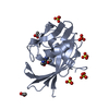
| ||||||||
| 2 | 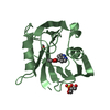
| ||||||||
| Unit cell |
|
-Components
-Protein , 1 types, 2 molecules AB
| #1: Protein | Mass: 18253.736 Da / Num. of mol.: 2 Source method: isolated from a genetically manipulated source Source: (gene. exp.) Homo sapiens (human) / Gene: NUDT1, MTH1 / Variant: ISOFORM P18 / Plasmid: PET-28A / Production host:  Escherichia coli (E. coli) / Strain (production host): ROSETTA PHAGE RESISTANT Escherichia coli (E. coli) / Strain (production host): ROSETTA PHAGE RESISTANTReferences: UniProt: P36639, 8-oxo-dGTP diphosphatase, 2-hydroxy-dATP diphosphatase |
|---|
-Non-polymers , 5 types, 111 molecules 


| #2: Chemical |  Mass: 190.199 Da / Num. of mol.: 2 / Source method: obtained synthetically / Formula: C10H10N2O2 / Feature type: SUBJECT OF INVESTIGATION Mass: 190.199 Da / Num. of mol.: 2 / Source method: obtained synthetically / Formula: C10H10N2O2 / Feature type: SUBJECT OF INVESTIGATION#3: Chemical | ChemComp-SO4 / Sulfate Mass: 96.063 Da / Num. of mol.: 6 / Source method: obtained synthetically / Formula: SO4 Mass: 96.063 Da / Num. of mol.: 6 / Source method: obtained synthetically / Formula: SO4#4: Chemical | Acetate Mass: 59.044 Da / Num. of mol.: 3 / Source method: obtained synthetically / Formula: C2H3O2 Mass: 59.044 Da / Num. of mol.: 3 / Source method: obtained synthetically / Formula: C2H3O2#5: Chemical | ChemComp-GOL / | Glycerol Mass: 92.094 Da / Num. of mol.: 1 / Source method: obtained synthetically / Formula: C3H8O3 Mass: 92.094 Da / Num. of mol.: 1 / Source method: obtained synthetically / Formula: C3H8O3#6: Water | ChemComp-HOH / | WaterMass: 18.015 Da / Num. of mol.: 99 / Source method: isolated from a natural source / Formula: H2O |
|---|
-Experimental details
-Experiment
| Experiment | Method: X-RAY DIFFRACTION / Number of used crystals: 1 |
|---|
- Sample preparation
| Crystal | Density Matthews: 2.32 Å3/Da / Density % sol: 47.02 % |
|---|---|
| Crystal grow | Temperature: 277 K / Method: vapor diffusion, sitting drop / pH: 3.6 Details: 27% PEG6000, 0.3M Lithium sulfate, 0.1M sodium acetate pH=3.6 |
-Data collection
| Diffraction | Mean temperature: 100 K |
|---|---|
| Diffraction source | Source: SYNCHROTRON / Site: ESRF  / Beamline: ID23-1 / Wavelength: 0.972 Å / Beamline: ID23-1 / Wavelength: 0.972 Å |
| Detector | Type: DECTRIS PILATUS 6M / Detector: PIXEL / Date: Mar 3, 2017 |
| Radiation | Protocol: SINGLE WAVELENGTH / Monochromatic (M) / Laue (L): M / Scattering type: x-ray |
| Radiation wavelength | Wavelength: 0.972 Å / Relative weight: 1 |
| Reflection | Resolution: 2→48.63 Å / Num. obs: 23476 / % possible obs: 98.9 % / Redundancy: 4.97 % / CC1/2: 0.999 / Rmerge(I) obs: 0.041 / Rrim(I) all: 0.046 / Net I/σ(I): 24.08 |
| Reflection shell | Resolution: 2→2.11 Å / Redundancy: 4.7 % / Rmerge(I) obs: 0.213 / Mean I/σ(I) obs: 8.14 / Num. unique obs: 3641 / CC1/2: 0.975 / Rrim(I) all: 0.24 / % possible all: 96.4 |
- Processing
| Software |
| ||||||||||||||||||||||||||||||||||||||||||||||||||||||||||||||||||||||||||||||||||||||||||||||||||||||||||||||||||||||||||||||||||||||||||||||||||||||||||||||||||||||||||||||||||||||
|---|---|---|---|---|---|---|---|---|---|---|---|---|---|---|---|---|---|---|---|---|---|---|---|---|---|---|---|---|---|---|---|---|---|---|---|---|---|---|---|---|---|---|---|---|---|---|---|---|---|---|---|---|---|---|---|---|---|---|---|---|---|---|---|---|---|---|---|---|---|---|---|---|---|---|---|---|---|---|---|---|---|---|---|---|---|---|---|---|---|---|---|---|---|---|---|---|---|---|---|---|---|---|---|---|---|---|---|---|---|---|---|---|---|---|---|---|---|---|---|---|---|---|---|---|---|---|---|---|---|---|---|---|---|---|---|---|---|---|---|---|---|---|---|---|---|---|---|---|---|---|---|---|---|---|---|---|---|---|---|---|---|---|---|---|---|---|---|---|---|---|---|---|---|---|---|---|---|---|---|---|---|---|---|
| Refinement | Method to determine structure: MOLECULAR REPLACEMENT Starting model: 5NHY Resolution: 2→48.63 Å / Cor.coef. Fo:Fc: 0.955 / Cor.coef. Fo:Fc free: 0.939 / SU B: 4.762 / SU ML: 0.128 / Cross valid method: THROUGHOUT / ESU R: 0.194 / ESU R Free: 0.167 / Details: HYDROGENS HAVE BEEN ADDED IN THE RIDING POSITIONS
| ||||||||||||||||||||||||||||||||||||||||||||||||||||||||||||||||||||||||||||||||||||||||||||||||||||||||||||||||||||||||||||||||||||||||||||||||||||||||||||||||||||||||||||||||||||||
| Solvent computation | Ion probe radii: 0.8 Å / Shrinkage radii: 0.8 Å / VDW probe radii: 1.2 Å | ||||||||||||||||||||||||||||||||||||||||||||||||||||||||||||||||||||||||||||||||||||||||||||||||||||||||||||||||||||||||||||||||||||||||||||||||||||||||||||||||||||||||||||||||||||||
| Displacement parameters | Biso mean: 30.722 Å2
| ||||||||||||||||||||||||||||||||||||||||||||||||||||||||||||||||||||||||||||||||||||||||||||||||||||||||||||||||||||||||||||||||||||||||||||||||||||||||||||||||||||||||||||||||||||||
| Refinement step | Cycle: 1 / Resolution: 2→48.63 Å
| ||||||||||||||||||||||||||||||||||||||||||||||||||||||||||||||||||||||||||||||||||||||||||||||||||||||||||||||||||||||||||||||||||||||||||||||||||||||||||||||||||||||||||||||||||||||
| Refine LS restraints |
|