 Movie
Movie Controller

[English] 日本語
 Yorodumi
- PDB-6ezc: Crystal Structure of human tRNA-dihydrouridine(20) synthase catal... -
+ Open data

- Basic information
| Entry | Database: PDB / ID: 6ezc | |||||||||
|---|---|---|---|---|---|---|---|---|---|---|
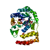
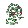
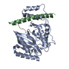
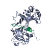
| Title | Crystal Structure of human tRNA-dihydrouridine(20) synthase catalytic domain E294K Q305K double mutant | |||||||||
 Components Components | tRNA-dihydrouridine(20) synthase [NAD(P)+]-like | |||||||||
 Keywords Keywords |  FLAVOPROTEIN / RNA BINDING PROTEIN / TRNA PROCESSING / OXIDOREDUCTASE FLAVOPROTEIN / RNA BINDING PROTEIN / TRNA PROCESSING / OXIDOREDUCTASE | |||||||||
| Function / homology |  Function and homology information Function and homology informationtRNA-dihydrouridine20 synthase [NAD(P)+] / tRNA-dihydrouridine20 synthase activity / tRNA dihydrouridine synthesis / tRNA dihydrouridine synthase activity / tRNA modification in the nucleus and cytosol / protein kinase inhibitor activity / antiviral innate immune response / NADPH binding / PKR-mediated signaling / double-stranded RNA binding ...tRNA-dihydrouridine20 synthase [NAD(P)+] / tRNA-dihydrouridine20 synthase activity / tRNA dihydrouridine synthesis / tRNA dihydrouridine synthase activity / tRNA modification in the nucleus and cytosol / protein kinase inhibitor activity / antiviral innate immune response / NADPH binding / PKR-mediated signaling / double-stranded RNA binding / FMN binding / flavin adenine dinucleotide binding / tRNA binding / endoplasmic reticulum / cytosol / cytoplasmSimilarity search - Function | |||||||||
| Biological species |  Homo sapiens (human) Homo sapiens (human) | |||||||||
| Method | X-RAY DIFFRACTION / SYNCHROTRON / MOLECULAR REPLACEMENT / Resolution: 2 Å | |||||||||
 Authors Authors | Bou-Nader, C. / Bregeon, D. / Vincent, G. / Fontecave, M. / Hamdane, D. | |||||||||
| Funding support |  France, 2items France, 2items
| |||||||||
 Citation Citation | Journal: Biochemistry / Year: 2018 Title: Electrostatic Potential in the tRNA Binding Evolution of Dihydrouridine Synthases. Authors: Bou-Nader, C. / Bregeon, D. / Pecqueur, L. / Fontecave, M. / Hamdane, D. | |||||||||
| History |
|
- Structure visualization
| Structure viewer | Molecule: MolmilJmol/JSmol |
|---|
- Downloads & links
-Download
| PDBx/mmCIF format | 6ezc.cif.gz | 148.5 KB | Display | PDBx/mmCIF format |
|---|---|---|---|---|
| PDB format | pdb6ezc.ent.gz | 113.2 KB | Display | PDB format |
| PDBx/mmJSON format | 6ezc.json.gz | Tree view | PDBx/mmJSON format | |
| Others |  Other downloads Other downloads |
-Validation report
| Arichive directory | https://data.pdbj.org/pub/pdb/validation_reports/ez/6ezcftp://data.pdbj.org/pub/pdb/validation_reports/ez/6ezc | HTTPS FTP |
|---|
-Related structure data
| Related structure data |  6ezaC  6ezbC  4xp7S C: citing same article ( S: Starting model for refinement |
|---|---|
| Similar structure data |


-Links
 PDBj
PDBj
- Assembly
| Deposited unit | 
| ||||||||
|---|---|---|---|---|---|---|---|---|---|
| 1 |
| ||||||||
| Unit cell |
|
-Components
-Protein , 1 types, 1 molecules A
| #1: Protein | Mass: 36806.477 Da / Num. of mol.: 1 Source method: isolated from a genetically manipulated source Source: (gene. exp.) Homo sapiens (human) / Gene: DUS2, DUS2L / Production host:  Escherichia coli (E. coli) / Variant (production host): pLysS Escherichia coli (E. coli) / Variant (production host): pLysSReferences: UniProt: Q9NX74, Oxidoreductases; Acting on the CH-CH group of donors; With NAD+ or NADP+ as acceptor |
|---|
-Non-polymers , 6 types, 240 molecules 


| #2: Chemical | ChemComp-FMN / Flavin mononucleotide Mass: 456.344 Da / Num. of mol.: 1 / Source method: obtained synthetically / Formula: C17H21N4O9P Mass: 456.344 Da / Num. of mol.: 1 / Source method: obtained synthetically / Formula: C17H21N4O9P | ||||||||
|---|---|---|---|---|---|---|---|---|---|
| #3: Chemical | ChemComp-SO4 / Sulfate Mass: 96.063 Da / Num. of mol.: 5 / Source method: obtained synthetically / Formula: SO4 Mass: 96.063 Da / Num. of mol.: 5 / Source method: obtained synthetically / Formula: SO4#4: Chemical | ChemComp-CL / | Chloride Mass: 35.453 Da / Num. of mol.: 1 / Source method: obtained synthetically / Formula: Cl Mass: 35.453 Da / Num. of mol.: 1 / Source method: obtained synthetically / Formula: Cl#5: Chemical | Glycerol Mass: 92.094 Da / Num. of mol.: 2 / Source method: obtained synthetically / Formula: C3H8O3 Mass: 92.094 Da / Num. of mol.: 2 / Source method: obtained synthetically / Formula: C3H8O3#6: Chemical | ChemComp-PGE / | Polyethylene glycol Mass: 150.173 Da / Num. of mol.: 1 / Source method: isolated from a natural source / Formula: C6H14O4 Mass: 150.173 Da / Num. of mol.: 1 / Source method: isolated from a natural source / Formula: C6H14O4#7: Water | ChemComp-HOH / | WaterMass: 18.015 Da / Num. of mol.: 230 / Source method: isolated from a natural source / Formula: H2O |
-Experimental details
-Experiment
| Experiment | Method: X-RAY DIFFRACTION / Number of used crystals: 1 |
|---|
- Sample preparation
| Crystal | Density Matthews: 2.66 Å3/Da / Density % sol: 53.8 % |
|---|---|
| Crystal grow | Temperature: 292 K / Method: vapor diffusion, hanging drop / pH: 5 Details: 30% PEG 2000 MME 200 mM ammonium sulfate 50 mM sodium acetate |
-Data collection
| Diffraction | Mean temperature: 100 K |
|---|---|
| Diffraction source | Source: SYNCHROTRON / Site: SOLEIL / Beamline: PROXIMA 2 / Wavelength: 0.980066 Å |
| Detector | Type: DECTRIS EIGER X 9M / Detector: PIXEL / Date: Sep 3, 2016 |
| Radiation | Protocol: SINGLE WAVELENGTH / Monochromatic (M) / Laue (L): M / Scattering type: x-ray |
| Radiation wavelength | Wavelength: 0.980066 Å / Relative weight: 1 |
| Reflection | Resolution: 2→43.17 Å / Num. obs: 22677 / % possible obs: 89.9 % / Redundancy: 9.3 % / CC1/2: 0.997 / Rmerge(I) obs: 0.161 / Rrim(I) all: 0.17 / Net I/σ(I): 10.1 |
| Reflection shell | Resolution: 2→2.11 Å / Redundancy: 8.9 % / Rmerge(I) obs: 2.521 / Mean I/σ(I) obs: 1.1 / Num. unique obs: 1135 / CC1/2: 0.413 / Rrim(I) all: 2.665 / % possible all: 43.5 |
- Processing
| Software |
| ||||||||||||||||||||||||||||||||||||||||||||||||||||||||||||||||||||||||||||||||||||||||||||||||||||||||||||||||||
|---|---|---|---|---|---|---|---|---|---|---|---|---|---|---|---|---|---|---|---|---|---|---|---|---|---|---|---|---|---|---|---|---|---|---|---|---|---|---|---|---|---|---|---|---|---|---|---|---|---|---|---|---|---|---|---|---|---|---|---|---|---|---|---|---|---|---|---|---|---|---|---|---|---|---|---|---|---|---|---|---|---|---|---|---|---|---|---|---|---|---|---|---|---|---|---|---|---|---|---|---|---|---|---|---|---|---|---|---|---|---|---|---|---|---|---|
| Refinement | Method to determine structure: MOLECULAR REPLACEMENT Starting model: 4XP7 Resolution: 2→42.33 Å / Cor.coef. Fo:Fc: 0.945 / Cor.coef. Fo:Fc free: 0.913 / SU R Cruickshank DPI: 0.183 / Cross valid method: THROUGHOUT / σ(F): 0 / SU R Blow DPI: 0.2 / SU Rfree Blow DPI: 0.169 / SU Rfree Cruickshank DPI: 0.162
| ||||||||||||||||||||||||||||||||||||||||||||||||||||||||||||||||||||||||||||||||||||||||||||||||||||||||||||||||||
| Displacement parameters | Biso mean: 39.49 Å2
| ||||||||||||||||||||||||||||||||||||||||||||||||||||||||||||||||||||||||||||||||||||||||||||||||||||||||||||||||||
| Refine analyze | Luzzati coordinate error obs: 0.24 Å | ||||||||||||||||||||||||||||||||||||||||||||||||||||||||||||||||||||||||||||||||||||||||||||||||||||||||||||||||||
| Refinement step | Cycle: 1 / Resolution: 2→42.33 Å
| ||||||||||||||||||||||||||||||||||||||||||||||||||||||||||||||||||||||||||||||||||||||||||||||||||||||||||||||||||
| Refine LS restraints |
| ||||||||||||||||||||||||||||||||||||||||||||||||||||||||||||||||||||||||||||||||||||||||||||||||||||||||||||||||||
| LS refinement shell | Resolution: 2→2.06 Å / Total num. of bins used: 50
| ||||||||||||||||||||||||||||||||||||||||||||||||||||||||||||||||||||||||||||||||||||||||||||||||||||||||||||||||||
| Refinement TLS params. | Method: refined / Origin x: 19.7569 Å / Origin y: -1.8799 Å / Origin z: -12.6109 Å
| ||||||||||||||||||||||||||||||||||||||||||||||||||||||||||||||||||||||||||||||||||||||||||||||||||||||||||||||||||
| Refinement TLS group | Selection details: { A|* } |
