 Movie
Movie Controller

[English] 日本語
 Yorodumi
- PDB-6eq0: Structure of the periplasmic binding protein (PBP) MelB (atu4661)... -
+ Open data

- Basic information
| Entry | Database: PDB / ID: 6eq0 | ||||||
|---|---|---|---|---|---|---|---|
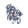
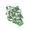
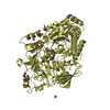
| Title | Structure of the periplasmic binding protein (PBP) MelB (atu4661) in complex with galactose from agrobacterium tumefacien C58 | ||||||
 Components Components | Periplasmic alpha-galactoside-binding protein | ||||||
 Keywords Keywords |  TRANSPORT PROTEIN / Protein transport associated to ABC transporter TRANSPORT PROTEIN / Protein transport associated to ABC transporter | ||||||
| Function / homology | Solute-binding protein family 5 domain / Solute-binding protein family 5 / Bacterial extracellular solute-binding proteins, family 5 Middle / periplasmic space / alpha-D-galactopyranose / DI(HYDROXYETHYL)ETHER / Periplasmic alpha-galactoside-binding protein Function and homology information Function and homology information | ||||||
| Biological species |  Rhizobium radiobacter (bacteria) Rhizobium radiobacter (bacteria) | ||||||
| Method | X-RAY DIFFRACTION / SYNCHROTRON / MOLECULAR REPLACEMENT / Resolution: 2.45 Å | ||||||
 Authors Authors | Vigouroux, A. / Morera, S. | ||||||
| Funding support |  France, 1items France, 1items
| ||||||
 Citation Citation | Journal: J. Biol. Chem. / Year: 2018 Title: The plant defense signal galactinol is specifically used as a nutrient by the bacterial pathogenAgrobacterium fabrum. Authors: Meyer, T. / Vigouroux, A. / Aumont-Nicaise, M. / Comte, G. / Vial, L. / Lavire, C. / Morera, S. | ||||||
| History |
|
- Structure visualization
| Structure viewer | Molecule: MolmilJmol/JSmol |
|---|
- Downloads & links
-Download
| PDBx/mmCIF format | 6eq0.cif.gz | 549.2 KB | Display | PDBx/mmCIF format |
|---|---|---|---|---|
| PDB format | pdb6eq0.ent.gz | 455.3 KB | Display | PDB format |
| PDBx/mmJSON format | 6eq0.json.gz | Tree view | PDBx/mmJSON format | |
| Others |  Other downloads Other downloads |
-Validation report
| Arichive directory | https://data.pdbj.org/pub/pdb/validation_reports/eq/6eq0ftp://data.pdbj.org/pub/pdb/validation_reports/eq/6eq0 | HTTPS FTP |
|---|
-Related structure data
| Related structure data |  6epyC  6epzSC  6eq1C  6eq8C C: citing same article ( S: Starting model for refinement |
|---|---|
| Similar structure data |


-Links
 PDBj
PDBj- Assembly
| Deposited unit | 
| ||||||||
|---|---|---|---|---|---|---|---|---|---|
| 1 | 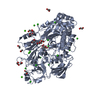
| ||||||||
| 2 | 
| ||||||||
| Unit cell |
|
-Components
-Protein / Sugars , 2 types, 4 molecules AB
| #1: Protein | Mass: 76359.602 Da / Num. of mol.: 2 Source method: isolated from a genetically manipulated source Source: (gene. exp.) Rhizobium radiobacter (bacteria) / Gene: SY94_4618 / Production host: Escherichia coli (E. coli) / References: UniProt: A0A083ZM57#2: Sugar | Galactose Type: D-saccharide, alpha linking / Mass: 180.156 Da / Num. of mol.: 2 Type: D-saccharide, alpha linking / Mass: 180.156 Da / Num. of mol.: 2Source method: isolated from a genetically manipulated source Formula: C6H12O6 |
|---|
-Non-polymers , 5 types, 309 molecules 


| #3: Chemical | Diethylene glycol Mass: 106.120 Da / Num. of mol.: 2 / Source method: obtained synthetically / Formula: C4H10O3 Mass: 106.120 Da / Num. of mol.: 2 / Source method: obtained synthetically / Formula: C4H10O3#4: Chemical | ChemComp-EDO / Ethylene glycol Mass: 62.068 Da / Num. of mol.: 21 / Source method: obtained synthetically / Formula: C2H6O2 Mass: 62.068 Da / Num. of mol.: 21 / Source method: obtained synthetically / Formula: C2H6O2#5: Chemical | ChemComp-CL / Chloride Mass: 35.453 Da / Num. of mol.: 16 / Source method: obtained synthetically / Formula: Cl Mass: 35.453 Da / Num. of mol.: 16 / Source method: obtained synthetically / Formula: Cl#6: Chemical | ChemComp-CA /  Mass: 40.078 Da / Num. of mol.: 17 / Source method: obtained synthetically / Formula: Ca Mass: 40.078 Da / Num. of mol.: 17 / Source method: obtained synthetically / Formula: Ca#7: Water | ChemComp-HOH / | WaterMass: 18.015 Da / Num. of mol.: 253 / Source method: isolated from a natural source / Formula: H2O |
|---|
-Experimental details
-Experiment
| Experiment | Method: X-RAY DIFFRACTION / Number of used crystals: 1 |
|---|
- Sample preparation
| Crystal | Density Matthews: 2.23 Å3/Da / Density % sol: 44.86 % |
|---|---|
| Crystal grow | Temperature: 298 K / Method: vapor diffusion, hanging drop Details: 25% PEG 4000, 0.6 M NaCl, 0.2 M CaCl2 and 0.1 M Mes pH 6.5 |
-Data collection
| Diffraction | Mean temperature: 100 K |
|---|---|
| Diffraction source | Source: SYNCHROTRON / Site: SOLEIL / Beamline: PROXIMA 2 / Wavelength: 0.98 Å |
| Detector | Type: DECTRIS EIGER X 9M / Detector: PIXEL / Date: Jun 6, 2015 |
| Radiation | Protocol: SINGLE WAVELENGTH / Monochromatic (M) / Laue (L): M / Scattering type: x-ray |
| Radiation wavelength | Wavelength: 0.98 Å / Relative weight: 1 |
| Reflection | Resolution: 2.45→50 Å / Num. obs: 49700 / % possible obs: 99.2 % / Redundancy: 6.3 % / Biso Wilson estimate: 63.81 Å2 / CC1/2: 0.991 / Rsym value: 0.182 / Net I/σ(I): 9 |
| Reflection shell | Resolution: 2.45→2.59 Å / Mean I/σ(I) obs: 1.6 / CC1/2: 0.512 / Rsym value: 1 / % possible all: 95.7 |
- Processing
| Software |
| ||||||||||||||||||||||||||||||||||||||||||||||||||||||||||||||||||||||||||||||||||||||||||||||||||||||||||||||||||
|---|---|---|---|---|---|---|---|---|---|---|---|---|---|---|---|---|---|---|---|---|---|---|---|---|---|---|---|---|---|---|---|---|---|---|---|---|---|---|---|---|---|---|---|---|---|---|---|---|---|---|---|---|---|---|---|---|---|---|---|---|---|---|---|---|---|---|---|---|---|---|---|---|---|---|---|---|---|---|---|---|---|---|---|---|---|---|---|---|---|---|---|---|---|---|---|---|---|---|---|---|---|---|---|---|---|---|---|---|---|---|---|---|---|---|---|
| Refinement | Method to determine structure: MOLECULAR REPLACEMENT Starting model: 6EPZ Resolution: 2.45→46.51 Å / Cor.coef. Fo:Fc: 0.921 / Cor.coef. Fo:Fc free: 0.891 / Rfactor Rfree error: 0 / SU R Cruickshank DPI: 0.665 / Cross valid method: THROUGHOUT / σ(F): 0 / SU R Blow DPI: 0.598 / SU Rfree Blow DPI: 0.27 / SU Rfree Cruickshank DPI: 0.278
| ||||||||||||||||||||||||||||||||||||||||||||||||||||||||||||||||||||||||||||||||||||||||||||||||||||||||||||||||||
| Displacement parameters | Biso mean: 65.47 Å2
| ||||||||||||||||||||||||||||||||||||||||||||||||||||||||||||||||||||||||||||||||||||||||||||||||||||||||||||||||||
| Refine analyze | Luzzati coordinate error obs: 0.34 Å | ||||||||||||||||||||||||||||||||||||||||||||||||||||||||||||||||||||||||||||||||||||||||||||||||||||||||||||||||||
| Refinement step | Cycle: 1 / Resolution: 2.45→46.51 Å
| ||||||||||||||||||||||||||||||||||||||||||||||||||||||||||||||||||||||||||||||||||||||||||||||||||||||||||||||||||
| Refine LS restraints |
| ||||||||||||||||||||||||||||||||||||||||||||||||||||||||||||||||||||||||||||||||||||||||||||||||||||||||||||||||||
| LS refinement shell | Resolution: 2.45→2.51 Å / Rfactor Rfree error: 0 / Total num. of bins used: 20
| ||||||||||||||||||||||||||||||||||||||||||||||||||||||||||||||||||||||||||||||||||||||||||||||||||||||||||||||||||
| Refinement TLS params. | Method: refined / Refine-ID: X-RAY DIFFRACTION
| ||||||||||||||||||||||||||||||||||||||||||||||||||||||||||||||||||||||||||||||||||||||||||||||||||||||||||||||||||
| Refinement TLS group |
|
