 Movie
Movie Controller

[English] 日本語
 Yorodumi
- PDB-6eio: Crystal structure of an ice binding protein from an Antarctic Bio... -
+ Open data

- Basic information
| Entry | Database: PDB / ID: 6eio | ||||||
|---|---|---|---|---|---|---|---|
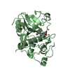
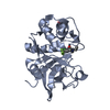
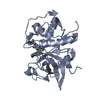
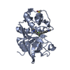
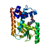
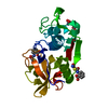
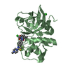
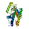
| Title | Crystal structure of an ice binding protein from an Antarctic Biological Consortium | ||||||
 Components Components | Antifreeze protein | ||||||
 Keywords Keywords | ANTIFREEZE PROTEIN / antifreeze / ice-binding | ||||||
| Function / homology | Ice-binding protein / Ice-binding-like / Antifreeze protein Function and homology information Function and homology information | ||||||
| Biological species |  uncultured bacterium (environmental samples) uncultured bacterium (environmental samples) | ||||||
| Method | X-RAY DIFFRACTION / SYNCHROTRON / MOLECULAR REPLACEMENT / Resolution: 0.84 Å | ||||||
 Authors Authors | Nardini, M. / Mangiagalli, M. / Nardone, V. / Bar Dolev, M. / Vena, V.F. / Sarusi, G. / Braslavsky, I. / Lotti, M. | ||||||
 Citation Citation | Journal: FEBS J. / Year: 2018 Title: Structure of a bacterial ice binding protein with two faces of interaction with ice. Authors: Mangiagalli, M. / Sarusi, G. / Kaleda, A. / Bar Dolev, M. / Nardone, V. / Vena, V.F. / Braslavsky, I. / Lotti, M. / Nardini, M. | ||||||
| History |
|
- Structure visualization
| Structure viewer | Molecule: MolmilJmol/JSmol |
|---|
- Downloads & links
-Download
| PDBx/mmCIF format | 6eio.cif.gz | 156.8 KB | Display | PDBx/mmCIF format |
|---|---|---|---|---|
| PDB format | pdb6eio.ent.gz | 123.9 KB | Display | PDB format |
| PDBx/mmJSON format | 6eio.json.gz | Tree view | PDBx/mmJSON format | |
| Others |  Other downloads Other downloads |
-Validation report
| Arichive directory | https://data.pdbj.org/pub/pdb/validation_reports/ei/6eioftp://data.pdbj.org/pub/pdb/validation_reports/ei/6eio | HTTPS FTP |
|---|
-Related structure data
| Related structure data | 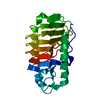 3wp9S S: Starting model for refinement |
|---|---|
| Similar structure data |


-Links
 PDBj
PDBj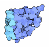
- Assembly
| Deposited unit | 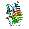
| ||||||||
|---|---|---|---|---|---|---|---|---|---|
| 1 |
| ||||||||
| Unit cell |
|
-Components
| #1: Protein | Mass: 24185.994 Da / Num. of mol.: 1 Source method: isolated from a genetically manipulated source Details: Ice-binding protein from the Antarctic ciliate Euplotes focardii and the associated bacterial consortium Source: (gene. exp.) uncultured bacterium (environmental samples)Production host: Escherichia coli (E. coli) / References: UniProt: A0A023J6X7 | ||
|---|---|---|---|
| #2: Chemical | ChemComp-GOL / Glycerol  Mass: 92.094 Da / Num. of mol.: 1 / Source method: obtained synthetically / Formula: C3H8O3 Mass: 92.094 Da / Num. of mol.: 1 / Source method: obtained synthetically / Formula: C3H8O3 | ||
| #3: Chemical | ChemComp-SO4 / Sulfate  Mass: 96.063 Da / Num. of mol.: 4 / Source method: obtained synthetically / Formula: SO4 Mass: 96.063 Da / Num. of mol.: 4 / Source method: obtained synthetically / Formula: SO4#4: Water | ChemComp-HOH / | Water Mass: 18.015 Da / Num. of mol.: 411 / Source method: isolated from a natural source / Formula: H2O Mass: 18.015 Da / Num. of mol.: 411 / Source method: isolated from a natural source / Formula: H2O |
-Experimental details
-Experiment
| Experiment | Method: X-RAY DIFFRACTION / Number of used crystals: 1 |
|---|
- Sample preparation
| Crystal | Density Matthews: 2.12 Å3/Da / Density % sol: 42 % |
|---|---|
| Crystal grow | Temperature: 293 K / Method: vapor diffusion, sitting drop / pH: 7.5 / Details: 2.2 M (NH4)2SO4, 0.1 M HEPES pH 7.5 |
-Data collection
| Diffraction | Mean temperature: 100 K |
|---|---|
| Diffraction source | Source: SYNCHROTRON / Site: ESRF  / Beamline: ID29 / Wavelength: 0.82656 Å / Beamline: ID29 / Wavelength: 0.82656 Å |
| Detector | Type: DECTRIS PILATUS3 S 6M / Detector: PIXEL / Date: Jun 20, 2016 |
| Radiation | Protocol: SINGLE WAVELENGTH / Monochromatic (M) / Laue (L): M / Scattering type: x-ray |
| Radiation wavelength | Wavelength: 0.82656 Å / Relative weight: 1 |
| Reflection | Resolution: 0.84→46.23 Å / Num. obs: 183816 / % possible obs: 94.5 % / Redundancy: 5.5 % / Rmerge(I) obs: 0.11 / Net I/σ(I): 10 |
| Reflection shell | Resolution: 0.84→0.89 Å / Redundancy: 3.1 % / Rmerge(I) obs: 0.58 / Mean I/σ(I) obs: 1.8 / Num. unique obs: 19237 / % possible all: 68.8 |
- Processing
| Software |
| |||||||||||||||||||||||||||||||||||||||||||||||||||||||||||||||||||||||||||||||||||||||||||||||||||||||||||||||||||||||||||||||||||||||||||||||||||||||||||||||||||||||||||||||||||||||||||||||||||||||||||||||||||||||||
|---|---|---|---|---|---|---|---|---|---|---|---|---|---|---|---|---|---|---|---|---|---|---|---|---|---|---|---|---|---|---|---|---|---|---|---|---|---|---|---|---|---|---|---|---|---|---|---|---|---|---|---|---|---|---|---|---|---|---|---|---|---|---|---|---|---|---|---|---|---|---|---|---|---|---|---|---|---|---|---|---|---|---|---|---|---|---|---|---|---|---|---|---|---|---|---|---|---|---|---|---|---|---|---|---|---|---|---|---|---|---|---|---|---|---|---|---|---|---|---|---|---|---|---|---|---|---|---|---|---|---|---|---|---|---|---|---|---|---|---|---|---|---|---|---|---|---|---|---|---|---|---|---|---|---|---|---|---|---|---|---|---|---|---|---|---|---|---|---|---|---|---|---|---|---|---|---|---|---|---|---|---|---|---|---|---|---|---|---|---|---|---|---|---|---|---|---|---|---|---|---|---|---|---|---|---|---|---|---|---|---|---|---|---|---|---|---|---|---|
| Refinement | Method to determine structure: MOLECULAR REPLACEMENT Starting model: 3WP9 Resolution: 0.84→46.226 Å / SU ML: 0.06 / Cross valid method: FREE R-VALUE / σ(F): 1.34 / Phase error: 11.26 / Details: anisotropic B-factors, H in protein atoms
| |||||||||||||||||||||||||||||||||||||||||||||||||||||||||||||||||||||||||||||||||||||||||||||||||||||||||||||||||||||||||||||||||||||||||||||||||||||||||||||||||||||||||||||||||||||||||||||||||||||||||||||||||||||||||
| Solvent computation | Shrinkage radii: 0.9 Å / VDW probe radii: 1.11 Å | |||||||||||||||||||||||||||||||||||||||||||||||||||||||||||||||||||||||||||||||||||||||||||||||||||||||||||||||||||||||||||||||||||||||||||||||||||||||||||||||||||||||||||||||||||||||||||||||||||||||||||||||||||||||||
| Refinement step | Cycle: LAST / Resolution: 0.84→46.226 Å
| |||||||||||||||||||||||||||||||||||||||||||||||||||||||||||||||||||||||||||||||||||||||||||||||||||||||||||||||||||||||||||||||||||||||||||||||||||||||||||||||||||||||||||||||||||||||||||||||||||||||||||||||||||||||||
| Refine LS restraints |
| |||||||||||||||||||||||||||||||||||||||||||||||||||||||||||||||||||||||||||||||||||||||||||||||||||||||||||||||||||||||||||||||||||||||||||||||||||||||||||||||||||||||||||||||||||||||||||||||||||||||||||||||||||||||||
| LS refinement shell |
|
