 Movie
Movie Controller


+ Open data

- Basic information
| Entry | Database: PDB / ID: 6efn | ||||||
|---|---|---|---|---|---|---|---|
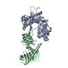
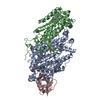
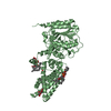
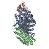
| Title | Structure of a RiPP maturase, SkfB | ||||||
 Components Components | Sporulation killing factor maturation protein SkfB | ||||||
 Keywords Keywords |  OXIDOREDUCTASE / S-Adenosylmethinine radical enzyme / metalloenzyme / natural product / RiPP / metal binding protein OXIDOREDUCTASE / S-Adenosylmethinine radical enzyme / metalloenzyme / natural product / RiPP / metal binding protein | ||||||
| Function / homology |  Function and homology informationOxidoreductases; Catalysing the reaction X-H + Y-H = X-Y; With other, known, physiological acceptors / bacteriocin biosynthetic process / 4 iron, 4 sulfur cluster binding / oxidoreductase activity / metal ion binding / cytoplasm Function and homology informationOxidoreductases; Catalysing the reaction X-H + Y-H = X-Y; With other, known, physiological acceptors / bacteriocin biosynthetic process / 4 iron, 4 sulfur cluster binding / oxidoreductase activity / metal ion binding / cytoplasmSimilarity search - Function | ||||||
| Biological species |  Bacillus subtilis (bacteria) Bacillus subtilis (bacteria) | ||||||
| Method | X-RAY DIFFRACTION / SYNCHROTRON / SAD / Resolution: 1.291 Å | ||||||
 Authors Authors | Grell, T.A.J. / Drennan, C.L. | ||||||
| Funding support |  United States, 1items United States, 1items
| ||||||
 Citation Citation | Journal: J.Biol.Chem. / Year: 2018 Title: Structure of a RiPP maturase, SkfB Authors: Grell, T.A.J. / Kincannon, W.M. / Bruender, N.A. / Blaesi, E.J. / Krebs, C. / Bandarian, V. / Drennan, C.L. | ||||||
| History |
|
- Structure visualization
| Structure viewer | Molecule: MolmilJmol/JSmol |
|---|
- Downloads & links
-Download
| PDBx/mmCIF format | 6efn.cif.gz | 182.3 KB | Display | PDBx/mmCIF format |
|---|---|---|---|---|
| PDB format | pdb6efn.ent.gz | 142.3 KB | Display | PDB format |
| PDBx/mmJSON format | 6efn.json.gz | Tree view | PDBx/mmJSON format | |
| Others |  Other downloads Other downloads |
-Validation report
| Arichive directory | https://data.pdbj.org/pub/pdb/validation_reports/ef/6efnftp://data.pdbj.org/pub/pdb/validation_reports/ef/6efn | HTTPS FTP |
|---|
-Related structure data
| Similar structure data |
|---|


-Links
 PDBj
PDBj


- Assembly
| Deposited unit | 
| ||||||||||||
|---|---|---|---|---|---|---|---|---|---|---|---|---|---|
| 1 | 
| ||||||||||||
| Unit cell |
| ||||||||||||
| Components on special symmetry positions |
|
-Components
-Protein , 1 types, 1 molecules A
| #1: Protein | Mass: 46002.230 Da / Num. of mol.: 1 Source method: isolated from a genetically manipulated source Source: (gene. exp.) Bacillus subtilis (strain 168) (bacteria)Strain: 168 / Gene: skfB, ybcP, ybcQ, BSU01920 / Production host: Escherichia coli (E. coli)References: UniProt: O31423, Oxidoreductases; Catalysing the reaction X-H + Y-H = X-Y; With other, known, physiological acceptors |
|---|
-Non-polymers , 5 types, 394 molecules 


| #2: Chemical | ChemComp-SAM / S-Adenosyl methionine Mass: 398.437 Da / Num. of mol.: 1 / Source method: obtained synthetically / Formula: C15H22N6O5S / Feature type: SUBJECT OF INVESTIGATION Mass: 398.437 Da / Num. of mol.: 1 / Source method: obtained synthetically / Formula: C15H22N6O5S / Feature type: SUBJECT OF INVESTIGATION |
|---|---|
| #3: Chemical | ChemComp-SF4 / Iron–sulfur cluster Mass: 351.640 Da / Num. of mol.: 1 / Source method: isolated from a natural source / Formula: Fe4S4 / Feature type: SUBJECT OF INVESTIGATION Mass: 351.640 Da / Num. of mol.: 1 / Source method: isolated from a natural source / Formula: Fe4S4 / Feature type: SUBJECT OF INVESTIGATION |
| #4: Chemical | ChemComp-FES / Iron–sulfur cluster Mass: 175.820 Da / Num. of mol.: 1 / Source method: obtained synthetically / Formula: Fe2S2 / Feature type: SUBJECT OF INVESTIGATION Mass: 175.820 Da / Num. of mol.: 1 / Source method: obtained synthetically / Formula: Fe2S2 / Feature type: SUBJECT OF INVESTIGATION |
| #5: Chemical | ChemComp-MG /  Mass: 24.305 Da / Num. of mol.: 1 / Source method: obtained synthetically / Formula: Mg Mass: 24.305 Da / Num. of mol.: 1 / Source method: obtained synthetically / Formula: Mg |
| #6: Water | ChemComp-HOH / WaterMass: 18.015 Da / Num. of mol.: 390 / Source method: isolated from a natural source / Formula: H2O |
-Experimental details
-Experiment
| Experiment | Method: X-RAY DIFFRACTION / Number of used crystals: 1 |
|---|
- Sample preparation
| Crystal | Density Matthews: 2.11 Å3/Da / Density % sol: 41.73 % |
|---|---|
| Crystal grow | Temperature: 294.15 K / Method: vapor diffusion, sitting drop / pH: 7.4 / Details: 0.25 M magnesium formate, 15% PEG3350 |
-Data collection
| Diffraction | Mean temperature: 100 K |
|---|---|
| Diffraction source | Source: SYNCHROTRON / Site: APS / Beamline: 24-ID-E / Wavelength: 0.9792 Å |
| Detector | Type: ADSC QUANTUM 315 / Detector: CCD / Date: Aug 4, 2013 |
| Radiation | Monochromator: Cryogenically-cooled single crystal Si(220) side bounce Protocol: SINGLE WAVELENGTH / Monochromatic (M) / Laue (L): M / Scattering type: x-ray |
| Radiation wavelength | Wavelength: 0.9792 Å / Relative weight: 1 |
| Reflection | Resolution: 1.291→42.574 Å / Num. obs: 92230 / % possible obs: 96.72 % / Redundancy: 5.6 % / Rsym value: 0.037 / Net I/σ(I): 43.6 |
| Reflection shell | Resolution: 1.291→1.34 Å / Redundancy: 2.6 % / Mean I/σ(I) obs: 1.8 / CC1/2: 0.729 / Rsym value: 0.499 / % possible all: 76.2 |
- Processing
| Software |
| |||||||||||||||||||||||||||||||||||||||||||||||||||||||||||||||||||||||||||||||||||||||||||||||||||||||||||||||||||||||||||||||||||||||||||||||||||||||||||||||||||||||||||||||||||||||||||||||||||||||||||||||||||||||||
|---|---|---|---|---|---|---|---|---|---|---|---|---|---|---|---|---|---|---|---|---|---|---|---|---|---|---|---|---|---|---|---|---|---|---|---|---|---|---|---|---|---|---|---|---|---|---|---|---|---|---|---|---|---|---|---|---|---|---|---|---|---|---|---|---|---|---|---|---|---|---|---|---|---|---|---|---|---|---|---|---|---|---|---|---|---|---|---|---|---|---|---|---|---|---|---|---|---|---|---|---|---|---|---|---|---|---|---|---|---|---|---|---|---|---|---|---|---|---|---|---|---|---|---|---|---|---|---|---|---|---|---|---|---|---|---|---|---|---|---|---|---|---|---|---|---|---|---|---|---|---|---|---|---|---|---|---|---|---|---|---|---|---|---|---|---|---|---|---|---|---|---|---|---|---|---|---|---|---|---|---|---|---|---|---|---|---|---|---|---|---|---|---|---|---|---|---|---|---|---|---|---|---|---|---|---|---|---|---|---|---|---|---|---|---|---|---|---|---|
| Refinement | Method to determine structure: SAD / Resolution: 1.291→42.574 Å / SU ML: 0.1 / Cross valid method: FREE R-VALUE / σ(F): 1.36 / Phase error: 20.6
| |||||||||||||||||||||||||||||||||||||||||||||||||||||||||||||||||||||||||||||||||||||||||||||||||||||||||||||||||||||||||||||||||||||||||||||||||||||||||||||||||||||||||||||||||||||||||||||||||||||||||||||||||||||||||
| Solvent computation | Shrinkage radii: 0.9 Å / VDW probe radii: 1.11 Å | |||||||||||||||||||||||||||||||||||||||||||||||||||||||||||||||||||||||||||||||||||||||||||||||||||||||||||||||||||||||||||||||||||||||||||||||||||||||||||||||||||||||||||||||||||||||||||||||||||||||||||||||||||||||||
| Refinement step | Cycle: LAST / Resolution: 1.291→42.574 Å
| |||||||||||||||||||||||||||||||||||||||||||||||||||||||||||||||||||||||||||||||||||||||||||||||||||||||||||||||||||||||||||||||||||||||||||||||||||||||||||||||||||||||||||||||||||||||||||||||||||||||||||||||||||||||||
| Refine LS restraints |
| |||||||||||||||||||||||||||||||||||||||||||||||||||||||||||||||||||||||||||||||||||||||||||||||||||||||||||||||||||||||||||||||||||||||||||||||||||||||||||||||||||||||||||||||||||||||||||||||||||||||||||||||||||||||||
| LS refinement shell |
|
