 Movie
Movie Controller

[English] 日本語
 Yorodumi
- PDB-6e9b: Bacteroides ovatus mixed-linkage glucan utilization locus (MLGUL)... -
+ Open data

- Basic information
| Entry | Database: PDB / ID: 6e9b | |||||||||
|---|---|---|---|---|---|---|---|---|---|---|
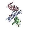
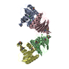
| Title | Bacteroides ovatus mixed-linkage glucan utilization locus (MLGUL) SGBP-B in complex with mixed-linkage heptasaccharide | |||||||||
 Components Components | Mixed-linkage glucan utilization locus (MLGUL) SGBP-B | |||||||||
 Keywords Keywords | SUGAR BINDING PROTEIN /  tetratricopeptide repeat / outer membrane protein / binding protein tetratricopeptide repeat / outer membrane protein / binding protein | |||||||||
| Function / homology | Immunoglobulin-like fold / PKD domain-containing protein Function and homology information Function and homology information | |||||||||
| Biological species |  Bacteroides ovatus (bacteria) Bacteroides ovatus (bacteria) | |||||||||
| Method | X-RAY DIFFRACTION / SYNCHROTRON / MOLECULAR REPLACEMENT / Resolution: 3.15 Å | |||||||||
 Authors Authors | Tamura, K. / Gardill, B.R. / Brumer, H. / Van Petegem, F. | |||||||||
 Citation Citation | Journal: Cell.Mol.Life Sci. / Year: 2019 Title: Surface glycan-binding proteins are essential for cereal beta-glucan utilization by the human gut symbiont Bacteroides ovatus. Authors: Tamura, K. / Foley, M.H. / Gardill, B.R. / Dejean, G. / Schnizlein, M. / Bahr, C.M.E. / Louise Creagh, A. / van Petegem, F. / Koropatkin, N.M. / Brumer, H. | |||||||||
| History |
|
- Structure visualization
| Structure viewer | Molecule: MolmilJmol/JSmol |
|---|
- Downloads & links
-Download
| PDBx/mmCIF format | 6e9b.cif.gz | 295.4 KB | Display | PDBx/mmCIF format |
|---|---|---|---|---|
| PDB format | pdb6e9b.ent.gz | 235.4 KB | Display | PDB format |
| PDBx/mmJSON format | 6e9b.json.gz | Tree view | PDBx/mmJSON format | |
| Others |  Other downloads Other downloads |
-Validation report
| Arichive directory | https://data.pdbj.org/pub/pdb/validation_reports/e9/6e9bftp://data.pdbj.org/pub/pdb/validation_reports/e9/6e9b | HTTPS FTP |
|---|
-Related structure data
| Related structure data |  6dmfC  6e57SC  6e60C  6e61C S: Starting model for refinement C: citing same article ( |
|---|---|
| Similar structure data |


-Links
 PDBj
PDBj- Assembly
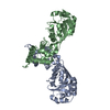
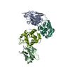
| Deposited unit | 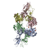
| ||||||||
|---|---|---|---|---|---|---|---|---|---|
| 1 | 
| ||||||||
| 2 | 
| ||||||||
| 3 | 
| ||||||||
| 4 | 
| ||||||||
| Unit cell |
|
-Components
| #1: Protein | Mass: 45678.328 Da / Num. of mol.: 4 Source method: isolated from a genetically manipulated source Source: (gene. exp.) Bacteroides ovatus (strain ATCC 8483 / DSM 1896 / JCM 5824 / NCTC 11153) (bacteria)Strain: ATCC 8483 / DSM 1896 / JCM 5824 / NCTC 11153 / Gene: BACOVA_02744 / Production host: Escherichia coli BL21(DE3) (bacteria) / Strain (production host): BL21(DE3) / References: UniProt: A7LY28#2: Polysaccharide | beta-D-glucopyranose-(1-4)-beta-D-glucopyranose-(1-3)-beta-D-glucopyranose-(1-4)-beta-D- ...beta-D-glucopyranose-(1-4)-beta-D-glucopyranose-(1-3)-beta-D-glucopyranose-(1-4)-beta-D-glucopyranose-(1-4)-beta-D-glucopyranose-(1-4)-beta-D-glucopyranose-(1-3)-beta-D-glucopyranose | / Mass: 1153.001 Da / Num. of mol.: 1Source method: isolated from a genetically manipulated source #3: Chemical | ChemComp-SO4 / Sulfate  Mass: 96.063 Da / Num. of mol.: 10 Mass: 96.063 Da / Num. of mol.: 10Source method: isolated from a genetically manipulated source Formula: SO4 #4: Water | ChemComp-HOH / | Water Mass: 18.015 Da / Num. of mol.: 67 / Source method: isolated from a natural source / Formula: H2O Mass: 18.015 Da / Num. of mol.: 67 / Source method: isolated from a natural source / Formula: H2O |
|---|
-Experimental details
-Experiment
| Experiment | Method: X-RAY DIFFRACTION / Number of used crystals: 1 |
|---|
- Sample preparation
| Crystal | Density Matthews: 3.9 Å3/Da / Density % sol: 71.59 % |
|---|---|
| Crystal grow | Temperature: 296 K / Method: vapor diffusion, hanging drop / pH: 4.2 Details: 0.2M lithium sulfate, 0.1M phosphate-citrate, 20% (w/v) PEG1000 Temp details: room temperature |
-Data collection
| Diffraction | Mean temperature: 100 K |
|---|---|
| Diffraction source | Source: SYNCHROTRON / Site: CLSI  / Beamline: 08B1-1 / Wavelength: 0.979 Å / Beamline: 08B1-1 / Wavelength: 0.979 Å |
| Detector | Type: RAYONIX MX300HE / Detector: CCD / Date: Feb 8, 2018 |
| Radiation | Protocol: SINGLE WAVELENGTH / Monochromatic (M) / Laue (L): M / Scattering type: x-ray |
| Radiation wavelength | Wavelength: 0.979 Å / Relative weight: 1 |
| Reflection | Resolution: 3.15→50 Å / Num. obs: 50094 / % possible obs: 99.8 % / Redundancy: 7.42 % / CC1/2: 0.999 / Net I/σ(I): 15.6 |
| Reflection shell | Resolution: 3.15→3.34 Å / Redundancy: 7.44 % / Mean I/σ(I) obs: 1.73 / Num. unique obs: 7894 / CC1/2: 0.651 / % possible all: 99.7 |
- Processing
| Software |
| ||||||||||||||||||||||||||||||||||||||||||||||||||||||||||||||||||||||||||||||||||||||||||||||||||||||||||||
|---|---|---|---|---|---|---|---|---|---|---|---|---|---|---|---|---|---|---|---|---|---|---|---|---|---|---|---|---|---|---|---|---|---|---|---|---|---|---|---|---|---|---|---|---|---|---|---|---|---|---|---|---|---|---|---|---|---|---|---|---|---|---|---|---|---|---|---|---|---|---|---|---|---|---|---|---|---|---|---|---|---|---|---|---|---|---|---|---|---|---|---|---|---|---|---|---|---|---|---|---|---|---|---|---|---|---|---|---|---|
| Refinement | Method to determine structure: MOLECULAR REPLACEMENT / Starting model: 6.0E+57 / Resolution: 3.15→34.33 Å / Cor.coef. Fo:Fc: 0.915 / Cor.coef. Fo:Fc free: 0.862 / SU R Cruickshank DPI: 1.087 / Cross valid method: THROUGHOUT / σ(F): 0 / SU R Blow DPI: 1.02 / SU Rfree Blow DPI: 0.398 / SU Rfree Cruickshank DPI: 0.407
| ||||||||||||||||||||||||||||||||||||||||||||||||||||||||||||||||||||||||||||||||||||||||||||||||||||||||||||
| Displacement parameters | Biso max: 207.63 Å2 / Biso mean: 100.53 Å2 / Biso min: 30.95 Å2
| ||||||||||||||||||||||||||||||||||||||||||||||||||||||||||||||||||||||||||||||||||||||||||||||||||||||||||||
| Refine analyze | Luzzati coordinate error obs: 0.46 Å | ||||||||||||||||||||||||||||||||||||||||||||||||||||||||||||||||||||||||||||||||||||||||||||||||||||||||||||
| Refinement step | Cycle: final / Resolution: 3.15→34.33 Å
| ||||||||||||||||||||||||||||||||||||||||||||||||||||||||||||||||||||||||||||||||||||||||||||||||||||||||||||
| Refine LS restraints |
| ||||||||||||||||||||||||||||||||||||||||||||||||||||||||||||||||||||||||||||||||||||||||||||||||||||||||||||
| LS refinement shell | Resolution: 3.15→3.18 Å / Rfactor Rfree error: 0 / Total num. of bins used: 50
|
