 Movie
Movie Controller


+ Open data

- Basic information
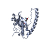
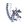
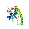
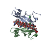
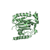
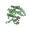
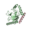
| Entry | Database: PDB / ID: 6e8h | ||||||
|---|---|---|---|---|---|---|---|
| Title | Legionella Longbeachae LeSH (Llo2327) | ||||||
 Components Components | LeSH (Llo2327) | ||||||
 Keywords Keywords | PEPTIDE BINDING PROTEIN /  Src homology 2 domain Src homology 2 domain | ||||||
| Function / homology | SH2 domain / Src homology 2 (SH2) domain profile. / SH2 domain / SH2 domain superfamily / SH2 domain-containing protein Function and homology information Function and homology information | ||||||
| Biological species | Legionella longbeachae serogroup 1 | ||||||
| Method | X-RAY DIFFRACTION / SAD / Resolution: 1.68 Å | ||||||
 Authors Authors | Kaneko, T. / Li, S.S.C. | ||||||
 Citation Citation | Journal: Nat Commun / Year: 2018 Title: Identification and characterization of a large family of superbinding bacterial SH2 domains. Authors: Kaneko, T. / Stogios, P.J. / Ruan, X. / Voss, C. / Evdokimova, E. / Skarina, T. / Chung, A. / Liu, X. / Li, L. / Savchenko, A. / Ensminger, A.W. / Li, S.S. | ||||||
| History |
|
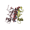
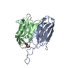
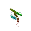
- Structure visualization
| Structure viewer | Molecule: MolmilJmol/JSmol |
|---|
- Downloads & links
-Download
| PDBx/mmCIF format | 6e8h.cif.gz | 100.6 KB | Display | PDBx/mmCIF format |
|---|---|---|---|---|
| PDB format | pdb6e8h.ent.gz | 63.6 KB | Display | PDB format |
| PDBx/mmJSON format | 6e8h.json.gz | Tree view | PDBx/mmJSON format | |
| Others |  Other downloads Other downloads |
-Validation report
| Arichive directory | https://data.pdbj.org/pub/pdb/validation_reports/e8/6e8hftp://data.pdbj.org/pub/pdb/validation_reports/e8/6e8h | HTTPS FTP |
|---|
-Related structure data


-Links
 PDBj
PDBj- Assembly
| Deposited unit | 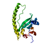
| ||||||||||||
|---|---|---|---|---|---|---|---|---|---|---|---|---|---|
| 1 |
| ||||||||||||
| Unit cell |
|
-Components
| #1: Protein | Mass: 19771.869 Da / Num. of mol.: 1 Source method: isolated from a genetically manipulated source Source: (gene. exp.)  Legionella longbeachae serogroup 1 (strain NSW150) (bacteria) Legionella longbeachae serogroup 1 (strain NSW150) (bacteria)Strain: NSW150 / Gene: LLO_2327 / Production host: Escherichia coli (E. coli) / References: UniProt: D3HJY4 |
|---|---|
| #2: Chemical | ChemComp-CL / Chloride  Mass: 35.453 Da / Num. of mol.: 1 / Source method: obtained synthetically / Formula: Cl Mass: 35.453 Da / Num. of mol.: 1 / Source method: obtained synthetically / Formula: Cl |
| #3: Water | ChemComp-HOH / Water Mass: 18.015 Da / Num. of mol.: 243 / Source method: isolated from a natural source / Formula: H2O Mass: 18.015 Da / Num. of mol.: 243 / Source method: isolated from a natural source / Formula: H2O |
-Experimental details
-Experiment
| Experiment | Method: X-RAY DIFFRACTION / Number of used crystals: 1 |
|---|
- Sample preparation
| Crystal | Density Matthews: 2.58 Å3/Da / Density % sol: 52.35 % |
|---|---|
| Crystal grow | Temperature: 278 K / Method: vapor diffusion / Details: Tris, PEG3000, sodium acetate |
-Data collection
| Diffraction | Mean temperature: 114 K |
|---|---|
| Diffraction source | Source: ROTATING ANODE / Type: RIGAKU RUH3R / Wavelength: 1.54 Å |
| Detector | Type: MAR scanner 345 mm plate / Detector: IMAGE PLATE / Date: Dec 1, 2012 |
| Radiation | Protocol: SINGLE WAVELENGTH / Monochromatic (M) / Laue (L): M / Scattering type: x-ray |
| Radiation wavelength | Wavelength: 1.54 Å / Relative weight: 1 |
| Reflection | Resolution: 1.66→19.63 Å / Num. obs: 22693 / % possible obs: 97.5 % / Redundancy: 55.7 % / Biso Wilson estimate: 21.17 Å2 / Rmerge(I) obs: 0.052 / Net I/σ(I): 68.2 |
| Reflection shell | Resolution: 1.66→1.76 Å / Redundancy: 50.5 % / Rmerge(I) obs: 0.284 / Mean I/σ(I) obs: 16.9 / Num. unique obs: 2832 / % possible all: 85 |
- Processing
| Software |
| |||||||||||||||||||||||||||||||||||||||||||||||||||||||||||||||||||||||||||||||||||||||||||||||||||||||||||||||||||||||||||||
|---|---|---|---|---|---|---|---|---|---|---|---|---|---|---|---|---|---|---|---|---|---|---|---|---|---|---|---|---|---|---|---|---|---|---|---|---|---|---|---|---|---|---|---|---|---|---|---|---|---|---|---|---|---|---|---|---|---|---|---|---|---|---|---|---|---|---|---|---|---|---|---|---|---|---|---|---|---|---|---|---|---|---|---|---|---|---|---|---|---|---|---|---|---|---|---|---|---|---|---|---|---|---|---|---|---|---|---|---|---|---|---|---|---|---|---|---|---|---|---|---|---|---|---|---|---|---|
| Refinement | Method to determine structure: SAD / Resolution: 1.68→19.63 Å / SU ML: 0.1958 / Cross valid method: FREE R-VALUE / σ(F): 1.34 / Phase error: 24.0777
| |||||||||||||||||||||||||||||||||||||||||||||||||||||||||||||||||||||||||||||||||||||||||||||||||||||||||||||||||||||||||||||
| Solvent computation | Shrinkage radii: 0.9 Å / VDW probe radii: 1.11 Å | |||||||||||||||||||||||||||||||||||||||||||||||||||||||||||||||||||||||||||||||||||||||||||||||||||||||||||||||||||||||||||||
| Displacement parameters | Biso mean: 29.79 Å2 | |||||||||||||||||||||||||||||||||||||||||||||||||||||||||||||||||||||||||||||||||||||||||||||||||||||||||||||||||||||||||||||
| Refinement step | Cycle: LAST / Resolution: 1.68→19.63 Å
| |||||||||||||||||||||||||||||||||||||||||||||||||||||||||||||||||||||||||||||||||||||||||||||||||||||||||||||||||||||||||||||
| Refine LS restraints |
| |||||||||||||||||||||||||||||||||||||||||||||||||||||||||||||||||||||||||||||||||||||||||||||||||||||||||||||||||||||||||||||
| LS refinement shell |
| |||||||||||||||||||||||||||||||||||||||||||||||||||||||||||||||||||||||||||||||||||||||||||||||||||||||||||||||||||||||||||||
| Refinement TLS params. | Method: refined / Refine-ID: X-RAY DIFFRACTION
| |||||||||||||||||||||||||||||||||||||||||||||||||||||||||||||||||||||||||||||||||||||||||||||||||||||||||||||||||||||||||||||
| Refinement TLS group |
|
