 Movie
Movie Controller

[English] 日本語
 Yorodumi
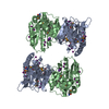
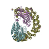
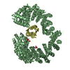
- PDB-6e8e: Crystal structure of the Escherichia coli sliding clamp-MutL complex. -
+ Open data

- Basic information
| Entry | Database: PDB / ID: 6e8e | ||||||
|---|---|---|---|---|---|---|---|
| Title | Crystal structure of the Escherichia coli sliding clamp-MutL complex. | ||||||
 Components Components | (Beta sliding clamp,DNA mismatch repair protein MutL) x 2 | ||||||
 Keywords Keywords |  DNA BINDING PROTEIN / Complex DNA BINDING PROTEIN / Complex | ||||||
| Function / homology |  Function and homology informationmismatch repair complex / Hda-beta clamp complex / bacterial-type DNA replication / replication inhibiting complex / mismatched DNA binding / DNA polymerase III complex / replisome / regulation of DNA-templated DNA replication initiation / DNA strand elongation involved in DNA replication / mismatch repair ...mismatch repair complex / Hda-beta clamp complex / bacterial-type DNA replication / replication inhibiting complex / mismatched DNA binding / DNA polymerase III complex / replisome / regulation of DNA-templated DNA replication initiation / DNA strand elongation involved in DNA replication / mismatch repair / error-prone translesion synthesis / negative regulation of DNA-templated DNA replication initiation / 3'-5' exonuclease activity / DNA-templated DNA replication / DNA-directed DNA polymerase activity / DNA damage response / ATP hydrolysis activity / protein homodimerization activity / DNA binding / ATP binding / identical protein binding / cytosol Function and homology informationmismatch repair complex / Hda-beta clamp complex / bacterial-type DNA replication / replication inhibiting complex / mismatched DNA binding / DNA polymerase III complex / replisome / regulation of DNA-templated DNA replication initiation / DNA strand elongation involved in DNA replication / mismatch repair ...mismatch repair complex / Hda-beta clamp complex / bacterial-type DNA replication / replication inhibiting complex / mismatched DNA binding / DNA polymerase III complex / replisome / regulation of DNA-templated DNA replication initiation / DNA strand elongation involved in DNA replication / mismatch repair / error-prone translesion synthesis / negative regulation of DNA-templated DNA replication initiation / 3'-5' exonuclease activity / DNA-templated DNA replication / DNA-directed DNA polymerase activity / DNA damage response / ATP hydrolysis activity / protein homodimerization activity / DNA binding / ATP binding / identical protein binding / cytosolSimilarity search - Function | ||||||
| Biological species |  Escherichia coli (E. coli)Escherichia coli O139:H28 (bacteria) Escherichia coli (E. coli)Escherichia coli O139:H28 (bacteria) | ||||||
| Method | X-RAY DIFFRACTION / SYNCHROTRON / MOLECULAR REPLACEMENT / Resolution: 2.25 Å | ||||||
 Authors Authors | Guarne, A. / Almawi, A.W. | ||||||
 Citation Citation | Journal: Nucleic Acids Res. / Year: 2019 Title: Binding of the regulatory domain of MutL to the sliding beta-clamp is species specific. Authors: Almawi, A.W. / Scotland, M.K. / Randall, J.R. / Liu, L. / Martin, H.K. / Sacre, L. / Shen, Y. / Pillon, M.C. / Simmons, L.A. / Sutton, M.D. / Guarne, A. | ||||||
| History |
|
- Structure visualization
| Structure viewer | Molecule: MolmilJmol/JSmol |
|---|
- Downloads & links
-Download
| PDBx/mmCIF format | 6e8e.cif.gz | 193.9 KB | Display | PDBx/mmCIF format |
|---|---|---|---|---|
| PDB format | pdb6e8e.ent.gz | 150.7 KB | Display | PDB format |
| PDBx/mmJSON format | 6e8e.json.gz | Tree view | PDBx/mmJSON format | |
| Others |  Other downloads Other downloads |
-Validation report
| Arichive directory | https://data.pdbj.org/pub/pdb/validation_reports/e8/6e8eftp://data.pdbj.org/pub/pdb/validation_reports/e8/6e8e | HTTPS FTP |
|---|
-Related structure data
| Related structure data |  6e8dC  1plqS  1x9zS S: Starting model for refinement C: citing same article ( |
|---|---|
| Similar structure data |


-Links
 PDBj
PDBj
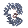
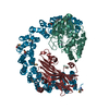
- Assembly
| Deposited unit | 
| ||||||||
|---|---|---|---|---|---|---|---|---|---|
| 1 | 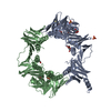
| ||||||||
| Unit cell |
|
-Components
| #1: Protein | Mass: 57098.820 Da / Num. of mol.: 1 Source method: isolated from a genetically manipulated source Source: (gene. exp.) Escherichia coli (strain K12) (bacteria), (gene. exp.) Escherichia coli O139:H28 (strain E24377A / ETEC) (bacteria)Strain: K12, E24377A / ETEC / Gene: dnaN, b3701, JW3678, mutL, EcE24377A_4728 / Production host: Escherichia coli BL21(DE3) (bacteria) / References: UniProt: P0A988, UniProt: A7ZV39 | ||||
|---|---|---|---|---|---|
| #2: Protein | Mass: 57082.820 Da / Num. of mol.: 1 Source method: isolated from a genetically manipulated source Source: (gene. exp.) Escherichia coli (strain K12) (bacteria), (gene. exp.) Escherichia coli O139:H28 (strain E24377A / ETEC) (bacteria)Strain: K12, E24377A / ETEC / Gene: dnaN, b3701, JW3678, mutL, EcE24377A_4728 / Production host: Escherichia coli BL21(DE3) (bacteria) / References: UniProt: P0A988, UniProt: A7ZV39 | ||||
| #3: Chemical | ChemComp-GOL / Glycerol  Mass: 92.094 Da / Num. of mol.: 6 / Source method: obtained synthetically / Formula: C3H8O3 Mass: 92.094 Da / Num. of mol.: 6 / Source method: obtained synthetically / Formula: C3H8O3#4: Chemical | ChemComp-SO4 / Sulfate  Mass: 96.063 Da / Num. of mol.: 5 / Source method: obtained synthetically / Formula: SO4 Mass: 96.063 Da / Num. of mol.: 5 / Source method: obtained synthetically / Formula: SO4#5: Water | ChemComp-HOH / | Water Mass: 18.015 Da / Num. of mol.: 191 / Source method: isolated from a natural source / Formula: H2O Mass: 18.015 Da / Num. of mol.: 191 / Source method: isolated from a natural source / Formula: H2O |
-Experimental details
-Experiment
| Experiment | Method: X-RAY DIFFRACTION / Number of used crystals: 1 |
|---|
- Sample preparation
| Crystal | Density Matthews: 2.51 Å3/Da / Density % sol: 50.98 % |
|---|---|
| Crystal grow | Temperature: 298 K / Method: vapor diffusion, hanging drop / Details: 100 mM Bis-Tris pH 5.5, 2 M ammonium sulfate. |
-Data collection
| Diffraction | Mean temperature: 100 K |
|---|---|
| Diffraction source | Source: SYNCHROTRON / Site: CLSI  / Beamline: 08ID-1 / Wavelength: 0.97951 Å / Beamline: 08ID-1 / Wavelength: 0.97951 Å |
| Detector | Type: DECTRIS PILATUS3 S 6M / Detector: PIXEL / Date: Jun 6, 2015 |
| Radiation | Protocol: SINGLE WAVELENGTH / Monochromatic (M) / Laue (L): M / Scattering type: x-ray |
| Radiation wavelength | Wavelength: 0.97951 Å / Relative weight: 1 |
| Reflection | Resolution: 2.07→46.83 Å / Num. obs: 53370 / % possible obs: 97.5 % / Redundancy: 4.4 % / CC1/2: 0.998 / Net I/σ(I): 11.5 |
| Reflection shell | Resolution: 2.25→2.32 Å / Num. unique all: 4976 / CC1/2: 0.356 |
- Processing
| Software |
| ||||||||||||||||||||||||||||||||||||||||||||||||||||||||||||||||||||||||||||||||||||
|---|---|---|---|---|---|---|---|---|---|---|---|---|---|---|---|---|---|---|---|---|---|---|---|---|---|---|---|---|---|---|---|---|---|---|---|---|---|---|---|---|---|---|---|---|---|---|---|---|---|---|---|---|---|---|---|---|---|---|---|---|---|---|---|---|---|---|---|---|---|---|---|---|---|---|---|---|---|---|---|---|---|---|---|---|---|
| Refinement | Method to determine structure: MOLECULAR REPLACEMENT Starting model: 1X9Z, 1PLQ Resolution: 2.25→46.83 Å / SU ML: 0.27 / Cross valid method: FREE R-VALUE / σ(F): 1.33 / Phase error: 24.05 / Stereochemistry target values: ML
| ||||||||||||||||||||||||||||||||||||||||||||||||||||||||||||||||||||||||||||||||||||
| Solvent computation | Shrinkage radii: 0.9 Å / VDW probe radii: 1.11 Å / Solvent model: FLAT BULK SOLVENT MODEL | ||||||||||||||||||||||||||||||||||||||||||||||||||||||||||||||||||||||||||||||||||||
| Refinement step | Cycle: LAST / Resolution: 2.25→46.83 Å
| ||||||||||||||||||||||||||||||||||||||||||||||||||||||||||||||||||||||||||||||||||||
| Refine LS restraints |
| ||||||||||||||||||||||||||||||||||||||||||||||||||||||||||||||||||||||||||||||||||||
| LS refinement shell |
|
