 Movie
Movie Controller

[English] 日本語
 Yorodumi
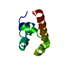
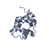
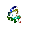
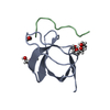
- PDB-6e4n: Structure of the T. brucei TbRGG2 RRM domain: apo R3 crystal form -
+ Open data

- Basic information
| Entry | Database: PDB / ID: 6e4n | ||||||
|---|---|---|---|---|---|---|---|
| Title | Structure of the T. brucei TbRGG2 RRM domain: apo R3 crystal form | ||||||
 Components Components | RNA-binding protein, putative | ||||||
 Keywords Keywords |  RNA BINDING PROTEIN / RRM / TbRGG2 / kRNA editing / RNA binding RNA BINDING PROTEIN / RRM / TbRGG2 / kRNA editing / RNA binding | ||||||
| Function / homology |  Function and homology information Function and homology informationmitochondrial mRNA processing / mitochondrial mRNA editing complex / cytoplasmic side of mitochondrial outer membrane / kinetoplast / RNA processing / mRNA binding / mitochondrion / nucleus / cytoplasmSimilarity search - Function | ||||||
| Biological species |  Trypanosoma brucei (eukaryote) Trypanosoma brucei (eukaryote) | ||||||
| Method | X-RAY DIFFRACTION / SYNCHROTRON / MOLECULAR REPLACEMENT / molecular replacement / Resolution: 1.801 Å | ||||||
 Authors Authors | Schumacher, M.A. | ||||||
 Citation Citation | Journal: Nucleic Acids Res. / Year: 2019 Title: The RRM of the kRNA-editing protein TbRGG2 uses multiple surfaces to bind and remodel RNA. Authors: Travis, B. / Shaw, P.L.R. / Liu, B. / Ravindra, K. / Iliff, H. / Al-Hashimi, H.M. / Schumacher, M.A. | ||||||
| History |
|
- Structure visualization
| Structure viewer | Molecule: MolmilJmol/JSmol |
|---|
- Downloads & links
-Download
| PDBx/mmCIF format | 6e4n.cif.gz | 42.2 KB | Display | PDBx/mmCIF format |
|---|---|---|---|---|
| PDB format | pdb6e4n.ent.gz | 27.7 KB | Display | PDB format |
| PDBx/mmJSON format | 6e4n.json.gz | Tree view | PDBx/mmJSON format | |
| Others |  Other downloads Other downloads |
-Validation report
| Arichive directory | https://data.pdbj.org/pub/pdb/validation_reports/e4/6e4nftp://data.pdbj.org/pub/pdb/validation_reports/e4/6e4n | HTTPS FTP |
|---|
-Related structure data
| Related structure data | 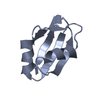 6e4oC  6e4pC  2m8dS S: Starting model for refinement C: citing same article ( |
|---|---|
| Similar structure data |


-Links
 PDBj
PDBj- Assembly
| Deposited unit | 
| ||||||||
|---|---|---|---|---|---|---|---|---|---|
| 1 |
| ||||||||
| Unit cell |
| ||||||||
| Components on special symmetry positions |
|
-Components
| #1: Protein | Mass: 7816.774 Da / Num. of mol.: 1 Source method: isolated from a genetically manipulated source Source: (gene. exp.) Trypanosoma brucei (eukaryote) / Production host:  Escherichia coli (E. coli) / References: UniProt: Q389P7 Escherichia coli (E. coli) / References: UniProt: Q389P7 |
|---|---|
| #2: Water | ChemComp-HOH / Water Mass: 18.015 Da / Num. of mol.: 93 / Source method: isolated from a natural source / Formula: H2O Mass: 18.015 Da / Num. of mol.: 93 / Source method: isolated from a natural source / Formula: H2O |
-Experimental details
-Experiment
| Experiment | Method: X-RAY DIFFRACTION / Number of used crystals: 1 |
|---|
- Sample preparation
| Crystal | Density Matthews: 2.54 Å3/Da / Density % sol: 51.58 % |
|---|---|
| Crystal grow | Temperature: 298 K / Method: vapor diffusion, hanging drop / Details: 1.4 M citrate, 0.1 M Bis-Tris pH 7 or HEPES |
-Data collection
| Diffraction | Mean temperature: 100 K |
|---|---|
| Diffraction source | Source: SYNCHROTRON / Site: ALS  / Beamline: 8.3.1 / Wavelength: 1 Å / Beamline: 8.3.1 / Wavelength: 1 Å |
| Detector | Type: ADSC QUANTUM 210r / Detector: CCD / Date: Feb 20, 2011 |
| Radiation | Protocol: SINGLE WAVELENGTH / Monochromatic (M) / Laue (L): M / Scattering type: x-ray |
| Radiation wavelength | Wavelength: 1 Å / Relative weight: 1 |
| Reflection | Resolution: 1.8→19.264 Å / Num. obs: 7045 / % possible obs: 99 % / Redundancy: 1.7 % / CC1/2: 0.994 / Rpim(I) all: 0.04 / Rsym value: 0.07 / Net I/σ(I): 11.3 |
| Reflection shell | Resolution: 1.8→1.99 Å / Redundancy: 1.7 % / CC1/2: 0.96 / Rpim(I) all: 0.161 / Rsym value: 0.249 / % possible all: 91.3 |
-Phasing
| Phasing | Method: molecular replacement |
|---|
- Processing
| Software |
| ||||||||||||||||||||||||||||||||||||||||
|---|---|---|---|---|---|---|---|---|---|---|---|---|---|---|---|---|---|---|---|---|---|---|---|---|---|---|---|---|---|---|---|---|---|---|---|---|---|---|---|---|---|
| Refinement | Method to determine structure: MOLECULAR REPLACEMENT Starting model: 2M8D Resolution: 1.801→19.264 Å / SU ML: 0.08 / Cross valid method: FREE R-VALUE / σ(F): 1.99 / Phase error: 17.98
| ||||||||||||||||||||||||||||||||||||||||
| Solvent computation | Shrinkage radii: 0.9 Å / VDW probe radii: 1.11 Å | ||||||||||||||||||||||||||||||||||||||||
| Displacement parameters | Biso max: 56.72 Å2 / Biso mean: 17.8 Å2 / Biso min: 3.27 Å2 | ||||||||||||||||||||||||||||||||||||||||
| Refinement step | Cycle: final / Resolution: 1.801→19.264 Å
| ||||||||||||||||||||||||||||||||||||||||
| Refine LS restraints |
| ||||||||||||||||||||||||||||||||||||||||
| LS refinement shell | Refine-ID: X-RAY DIFFRACTION / Rfactor Rfree error: 0 / Total num. of bins used: 2
| ||||||||||||||||||||||||||||||||||||||||
| Refinement TLS params. | Method: refined / Origin x: -13.8585 Å / Origin y: 2.0009 Å / Origin z: -5.1759 Å
| ||||||||||||||||||||||||||||||||||||||||
| Refinement TLS group |
|
