 Movie
Movie Controller


+ Open data

- Basic information
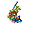
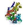
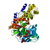
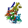
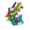
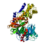
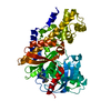
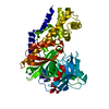
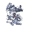
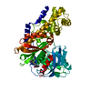
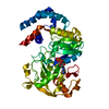
| Entry | Database: PDB / ID: 6e0e | ||||||
|---|---|---|---|---|---|---|---|
| Title | Crystal structure of Glucokinase in complex with compound 6 | ||||||
 Components Components | Glucokinase | ||||||
 Keywords Keywords | Transferase/Transferase Inhibitor / Glucokinase / glucokinase activator / structure-aided design / structure-based design / type II diabetes / Transferase-Transferase Inhibitor complex | ||||||
| Function / homology |  Function and homology information Function and homology informationDefective GCK causes maturity-onset diabetes of the young 2 (MODY2) / mannokinase activity / glucose sensor activity / regulation of potassium ion transport / hexokinase / fructokinase activity / carbohydrate phosphorylation / glucokinase activity / glucose catabolic process / glucose 6-phosphate metabolic process ...Defective GCK causes maturity-onset diabetes of the young 2 (MODY2) / mannokinase activity / glucose sensor activity / regulation of potassium ion transport / hexokinase / fructokinase activity / carbohydrate phosphorylation / glucokinase activity / glucose catabolic process / glucose 6-phosphate metabolic process / NADP metabolic process / Regulation of Glucokinase by Glucokinase Regulatory Protein / Defective TPR may confer susceptibility towards thyroid papillary carcinoma (TPC) / D-glucose binding / cellular response to leptin stimulus / calcium ion import / canonical glycolysis / Glycolysis / regulation of glycolytic process / intracellular glucose homeostasis / Regulation of gene expression in beta cells / regulation of insulin secretion / positive regulation of glycogen biosynthetic process / FOXO-mediated transcription of oxidative stress, metabolic and neuronal genes / negative regulation of gluconeogenesis / response to glucose / glycolytic process / positive regulation of insulin secretion / cellular response to insulin stimulus / glucose metabolic process / glucose homeostasis / mitochondrion / nucleoplasm / ATP binding / cytosolSimilarity search - Function | ||||||
| Biological species |  Homo sapiens (human) Homo sapiens (human) | ||||||
| Method | X-RAY DIFFRACTION / FOURIER SYNTHESIS / Resolution: 2.7 Å | ||||||
 Authors Authors | Hinklin, R.J. / Baer, B.R. / Boyd, S.A. / Chicarelli, M.D. / Condroski, K.R. / DeWolf, W.E. / Fischer, J. / Frank, M. / Hingorani, G.P. / Lee, P.A. ...Hinklin, R.J. / Baer, B.R. / Boyd, S.A. / Chicarelli, M.D. / Condroski, K.R. / DeWolf, W.E. / Fischer, J. / Frank, M. / Hingorani, G.P. / Lee, P.A. / Neitzel, N.A. / Pratt, S.A. / Singh, A. / Sullivan, F.X. / Turner, T. / Voegtli, W.C. / Wallace, E.M. / Williams, L. / Aicher, T.D. | ||||||
 Citation Citation | Journal: Bioorg.Med.Chem. / Year: 2020 Title: Discovery and preclinical development of AR453588 as an anti-diabetic glucokinase activator. Authors: Hinklin, R.J. / Baer, B.R. / Boyd, S.A. / Chicarelli, M.D. / Condroski, K.R. / DeWolf Jr., W.E. / Fischer, J. / Frank, M. / Hingorani, G.P. / Lee, P.A. / Neitzel, N.A. / Pratt, S.A. / Singh, ...Authors: Hinklin, R.J. / Baer, B.R. / Boyd, S.A. / Chicarelli, M.D. / Condroski, K.R. / DeWolf Jr., W.E. / Fischer, J. / Frank, M. / Hingorani, G.P. / Lee, P.A. / Neitzel, N.A. / Pratt, S.A. / Singh, A. / Sullivan, F.X. / Turner, T. / Voegtli, W.C. / Wallace, E.M. / Williams, L. / Aicher, T.D. | ||||||
| History |
|
- Structure visualization
| Structure viewer | Molecule: MolmilJmol/JSmol |
|---|
- Downloads & links
-Download
| PDBx/mmCIF format | 6e0e.cif.gz | 104.1 KB | Display | PDBx/mmCIF format |
|---|---|---|---|---|
| PDB format | pdb6e0e.ent.gz | 78.4 KB | Display | PDB format |
| PDBx/mmJSON format | 6e0e.json.gz | Tree view | PDBx/mmJSON format | |
| Others |  Other downloads Other downloads |
-Validation report
| Arichive directory | https://data.pdbj.org/pub/pdb/validation_reports/e0/6e0eftp://data.pdbj.org/pub/pdb/validation_reports/e0/6e0e | HTTPS FTP |
|---|
-Related structure data
-Links
 PDBj
PDBj


- Assembly
| Deposited unit | 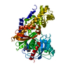
| ||||||||
|---|---|---|---|---|---|---|---|---|---|
| 1 |
| ||||||||
| Unit cell |
|
-Components
| #1: Protein | / Hexokinase type IV / HK IV / Hexokinase-4 / HK4 / Hexokinase-D Mass: 50264.035 Da / Num. of mol.: 1 Source method: isolated from a genetically manipulated source Source: (gene. exp.) Homo sapiens (human) / Gene: GCK / Production host:  Escherichia coli (E. coli) / References: UniProt: P35557, glucokinase Escherichia coli (E. coli) / References: UniProt: P35557, glucokinase |
|---|---|
| #2: Sugar | ChemComp-GLC / Glucose  Type: D-saccharide, alpha linking / Mass: 180.156 Da / Num. of mol.: 1 Type: D-saccharide, alpha linking / Mass: 180.156 Da / Num. of mol.: 1Source method: isolated from a genetically manipulated source Formula: C6H12O6 |
| #3: Chemical | ChemComp-HKM /   Mass: 308.358 Da / Num. of mol.: 1 / Source method: obtained synthetically / Formula: C16H12N4OS / Feature type: SUBJECT OF INVESTIGATION Mass: 308.358 Da / Num. of mol.: 1 / Source method: obtained synthetically / Formula: C16H12N4OS / Feature type: SUBJECT OF INVESTIGATION |
| #4: Water | ChemComp-HOH / Water Mass: 18.015 Da / Num. of mol.: 64 / Source method: isolated from a natural source / Formula: H2O Mass: 18.015 Da / Num. of mol.: 64 / Source method: isolated from a natural source / Formula: H2O |
-Experimental details
-Experiment
| Experiment | Method: X-RAY DIFFRACTION / Number of used crystals: 1 |
|---|
- Sample preparation
| Crystal | Density Matthews: 2.91 Å3/Da / Density % sol: 57.74 % |
|---|---|
| Crystal grow | Temperature: 293 K / Method: vapor diffusion, hanging drop / pH: 7 / Details: 0.1M Hepes pH7.0, 27% PEG1500 |
-Data collection
| Diffraction | Mean temperature: 100 K |
|---|---|
| Diffraction source | Source: ROTATING ANODE / Type: RIGAKU FR-E / Wavelength: 1.5418 Å |
| Detector | Type: RIGAKU RAXIS IV++ / Detector: IMAGE PLATE / Date: Oct 6, 2005 |
| Radiation | Protocol: SINGLE WAVELENGTH / Monochromatic (M) / Laue (L): M / Scattering type: x-ray |
| Radiation wavelength | Wavelength: 1.5418 Å / Relative weight: 1 |
| Reflection | Resolution: 2.7→28.95 Å / Num. obs: 16310 / % possible obs: 93.1 % / Redundancy: 5.5 % / Net I/σ(I): 10.7 |
| Reflection shell | Resolution: 2.7→2.85 Å |
- Processing
| Software |
| ||||||||||||||||||||||||||||||||||||||||||||||||||||||||||||||||||||||||||||||||||||||||||||||||||||||||||||||||||||||||||||||||||||||||||||||||||||||||||||||||||||||||||||||||||||||
|---|---|---|---|---|---|---|---|---|---|---|---|---|---|---|---|---|---|---|---|---|---|---|---|---|---|---|---|---|---|---|---|---|---|---|---|---|---|---|---|---|---|---|---|---|---|---|---|---|---|---|---|---|---|---|---|---|---|---|---|---|---|---|---|---|---|---|---|---|---|---|---|---|---|---|---|---|---|---|---|---|---|---|---|---|---|---|---|---|---|---|---|---|---|---|---|---|---|---|---|---|---|---|---|---|---|---|---|---|---|---|---|---|---|---|---|---|---|---|---|---|---|---|---|---|---|---|---|---|---|---|---|---|---|---|---|---|---|---|---|---|---|---|---|---|---|---|---|---|---|---|---|---|---|---|---|---|---|---|---|---|---|---|---|---|---|---|---|---|---|---|---|---|---|---|---|---|---|---|---|---|---|---|---|
| Refinement | Method to determine structure: FOURIER SYNTHESIS / Resolution: 2.7→28.95 Å / Cor.coef. Fo:Fc: 0.938 / Cor.coef. Fo:Fc free: 0.876 / SU B: 14.276 / SU ML: 0.279 / Cross valid method: THROUGHOUT / ESU R: 1.073 / ESU R Free: 0.376
| ||||||||||||||||||||||||||||||||||||||||||||||||||||||||||||||||||||||||||||||||||||||||||||||||||||||||||||||||||||||||||||||||||||||||||||||||||||||||||||||||||||||||||||||||||||||
| Solvent computation | Ion probe radii: 0.8 Å / Shrinkage radii: 0.8 Å / VDW probe radii: 1.2 Å | ||||||||||||||||||||||||||||||||||||||||||||||||||||||||||||||||||||||||||||||||||||||||||||||||||||||||||||||||||||||||||||||||||||||||||||||||||||||||||||||||||||||||||||||||||||||
| Displacement parameters | Biso mean: 55.307 Å2
| ||||||||||||||||||||||||||||||||||||||||||||||||||||||||||||||||||||||||||||||||||||||||||||||||||||||||||||||||||||||||||||||||||||||||||||||||||||||||||||||||||||||||||||||||||||||
| Refinement step | Cycle: 1 / Resolution: 2.7→28.95 Å
| ||||||||||||||||||||||||||||||||||||||||||||||||||||||||||||||||||||||||||||||||||||||||||||||||||||||||||||||||||||||||||||||||||||||||||||||||||||||||||||||||||||||||||||||||||||||
| Refine LS restraints |
|