 Movie
Movie Controller

[English] 日本語
 Yorodumi
- PDB-6dxt: Structure of USP5 zinc-finger ubiquitin binding domain co-crystal... -
+ Open data

- Basic information
| Entry | Database: PDB / ID: 6dxt | ||||||
|---|---|---|---|---|---|---|---|
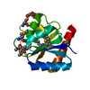
| Title | Structure of USP5 zinc-finger ubiquitin binding domain co-crystallized with 3-(5-phenyl-1,3,4-oxadiazol-2-yl)propanoate | ||||||
 Components Components | Ubiquitin carboxyl-terminal hydrolase 5 | ||||||
 Keywords Keywords |  HYDROLASE / DEUBIQUITINASE / USP5 / UBIQUITIN / STRUCTURAL GENOMICS CONSORTIUM / SGC HYDROLASE / DEUBIQUITINASE / USP5 / UBIQUITIN / STRUCTURAL GENOMICS CONSORTIUM / SGC | ||||||
| Function / homology |  Function and homology information Function and homology informationprotein K48-linked deubiquitination / negative regulation of ubiquitin-dependent protein catabolic process / Synthesis of active ubiquitin: roles of E1 and E2 enzymes / ubiquitin binding / positive regulation of proteasomal ubiquitin-dependent protein catabolic process / ubiquitinyl hydrolase 1 / cysteine-type deubiquitinase activity / lysosome / protein ubiquitination / Ub-specific processing proteases ...protein K48-linked deubiquitination / negative regulation of ubiquitin-dependent protein catabolic process / Synthesis of active ubiquitin: roles of E1 and E2 enzymes / ubiquitin binding / positive regulation of proteasomal ubiquitin-dependent protein catabolic process / ubiquitinyl hydrolase 1 / cysteine-type deubiquitinase activity / lysosome / protein ubiquitination / Ub-specific processing proteases / cysteine-type endopeptidase activity / proteolysis / zinc ion binding / nucleus / cytosolSimilarity search - Function | ||||||
| Biological species |  Homo sapiens (human) Homo sapiens (human) | ||||||
| Method | X-RAY DIFFRACTION / MOLECULAR REPLACEMENT / Resolution: 1.95 Å | ||||||
 Authors Authors | Mann, M.K. / Harding, R.J. / Ravichandran, M. / Ferreira de Freitas, R. / Franzoni, I. / Bountra, C. / Edwards, A.M. / Arrowsmith, C.M. / Schapira, M. / Structural Genomics Consortium (SGC) | ||||||
 Citation Citation | Journal: J.Med.Chem. / Year: 2019 Title: Discovery of Small Molecule Antagonists of the USP5 Zinc Finger Ubiquitin-Binding Domain. Authors: Mann, M.K. / Franzoni, I. / de Freitas, R.F. / Tempel, W. / Houliston, S. / Smith, L. / Vedadi, M. / Arrowsmith, C.H. / Harding, R.J. / Schapira, M. | ||||||
| History |
|
- Structure visualization
| Structure viewer | Molecule: MolmilJmol/JSmol |
|---|
- Downloads & links
-Download
| PDBx/mmCIF format | 6dxt.cif.gz | 64.9 KB | Display | PDBx/mmCIF format |
|---|---|---|---|---|
| PDB format | pdb6dxt.ent.gz | 45.3 KB | Display | PDB format |
| PDBx/mmJSON format | 6dxt.json.gz | Tree view | PDBx/mmJSON format | |
| Others |  Other downloads Other downloads |
-Validation report
| Arichive directory | https://data.pdbj.org/pub/pdb/validation_reports/dx/6dxtftp://data.pdbj.org/pub/pdb/validation_reports/dx/6dxt | HTTPS FTP |
|---|
-Related structure data
| Related structure data |  6dxhC  6nftC  6p9gC  2g43S S: Starting model for refinement C: citing same article ( |
|---|---|
| Similar structure data |


-Links
 PDBj
PDBj


- Assembly
| Deposited unit | 
| ||||||||
|---|---|---|---|---|---|---|---|---|---|
| 1 | 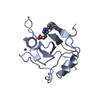
| ||||||||
| 2 | 
| ||||||||
| Unit cell |
|
-Components
-Protein , 1 types, 2 molecules AB
| #1: Protein | Mass: 13535.199 Da / Num. of mol.: 2 / Fragment: UNP residues 171-290 Source method: isolated from a genetically manipulated source Source: (gene. exp.) Homo sapiens (human) / Gene: USP5, ISOT / Plasmid: pET28-mhl / Production host:  Escherichia coli (E. coli) / Strain (production host): BL21 (DE3) codon plus / References: UniProt: P45974, ubiquitinyl hydrolase 1 Escherichia coli (E. coli) / Strain (production host): BL21 (DE3) codon plus / References: UniProt: P45974, ubiquitinyl hydrolase 1 |
|---|
-Non-polymers , 5 types, 177 molecules 


| #2: Chemical | ChemComp-HHY /  Mass: 218.209 Da / Num. of mol.: 1 / Source method: isolated from a natural source / Formula: C11H10N2O3 Mass: 218.209 Da / Num. of mol.: 1 / Source method: isolated from a natural source / Formula: C11H10N2O3 | ||||||
|---|---|---|---|---|---|---|---|
| #3: Chemical |  Mass: 62.068 Da / Num. of mol.: 2 / Source method: isolated from a natural source Mass: 62.068 Da / Num. of mol.: 2 / Source method: isolated from a natural source#4: Chemical |  Mass: 65.409 Da / Num. of mol.: 2 / Source method: isolated from a natural source / Formula: Zn Mass: 65.409 Da / Num. of mol.: 2 / Source method: isolated from a natural source / Formula: Zn#5: Chemical | Ethylene glycol Mass: 62.068 Da / Num. of mol.: 2 / Source method: isolated from a natural source / Formula: C2H6O2 Mass: 62.068 Da / Num. of mol.: 2 / Source method: isolated from a natural source / Formula: C2H6O2#6: Water | ChemComp-HOH / | WaterMass: 18.015 Da / Num. of mol.: 170 / Source method: isolated from a natural source / Formula: H2O |
-Experimental details
-Experiment
| Experiment | Method: X-RAY DIFFRACTION / Number of used crystals: 1 |
|---|
- Sample preparation
| Crystal | Density Matthews: 2.83 Å3/Da / Density % sol: 56.59 % / Mosaicity: 0 ° |
|---|---|
| Crystal grow | Temperature: 291 K / Method: vapor diffusion, sitting drop / pH: 7 Details: 1.5 M ammonium sulfate, 0.1 M bis-tris pH 7.0, 25% ethylene glycol (v/v), 1.1% DMSO (v/v) |
-Data collection
| Diffraction | Mean temperature: 100 K | ||||||||||||||||||||||||||||||
|---|---|---|---|---|---|---|---|---|---|---|---|---|---|---|---|---|---|---|---|---|---|---|---|---|---|---|---|---|---|---|---|
| Diffraction source | Source: ROTATING ANODE / Type: RIGAKU FR-E SUPERBRIGHT / Wavelength: 1.54178 Å | ||||||||||||||||||||||||||||||
| Detector | Type: RIGAKU SATURN A200 / Detector: CCD / Date: May 23, 2018 | ||||||||||||||||||||||||||||||
| Radiation | Protocol: SINGLE WAVELENGTH / Monochromatic (M) / Laue (L): M / Scattering type: x-ray | ||||||||||||||||||||||||||||||
| Radiation wavelength | Wavelength: 1.54178 Å / Relative weight: 1 | ||||||||||||||||||||||||||||||
| Reflection | Resolution: 1.95→30.05 Å / Num. obs: 20938 / % possible obs: 95.2 % / Redundancy: 3.8 % / CC1/2: 0.999 / Rmerge(I) obs: 0.038 / Rpim(I) all: 0.022 / Rrim(I) all: 0.044 / Net I/σ(I): 16.2 / Num. measured all: 80002 | ||||||||||||||||||||||||||||||
| Reflection shell | Diffraction-ID: 1
|
- Processing
| Software |
| ||||||||||||||||||||||||||||||||||||||||||||||||||||||||||||
|---|---|---|---|---|---|---|---|---|---|---|---|---|---|---|---|---|---|---|---|---|---|---|---|---|---|---|---|---|---|---|---|---|---|---|---|---|---|---|---|---|---|---|---|---|---|---|---|---|---|---|---|---|---|---|---|---|---|---|---|---|---|
| Refinement | Method to determine structure: MOLECULAR REPLACEMENT Starting model: pdbid 2G43 Resolution: 1.95→30.07 Å / Cor.coef. Fo:Fc: 0.945 / Cor.coef. Fo:Fc free: 0.918 / SU B: 3.155 / SU ML: 0.091 / Cross valid method: THROUGHOUT / σ(F): 0 / ESU R: 0.157 / ESU R Free: 0.152 / Stereochemistry target values: MAXIMUM LIKELIHOOD Details: Users of this crystal structure: verify our intepretion of the electron density. Additional ambiguous density is observed in the binding pocket of chain B, and is suspected to be low ...Details: Users of this crystal structure: verify our intepretion of the electron density. Additional ambiguous density is observed in the binding pocket of chain B, and is suspected to be low occupancy 3-(5-Phenyl-1,3,4-oxadiazol-2-yl)propanoate. LC-MS evaluation of the ligand shows a
| ||||||||||||||||||||||||||||||||||||||||||||||||||||||||||||
| Solvent computation | Ion probe radii: 0.8 Å / Shrinkage radii: 0.8 Å / VDW probe radii: 1.2 Å / Solvent model: MASK | ||||||||||||||||||||||||||||||||||||||||||||||||||||||||||||
| Displacement parameters | Biso max: 63.62 Å2 / Biso mean: 25.318 Å2 / Biso min: 12.74 Å2
| ||||||||||||||||||||||||||||||||||||||||||||||||||||||||||||
| Refinement step | Cycle: final / Resolution: 1.95→30.07 Å
| ||||||||||||||||||||||||||||||||||||||||||||||||||||||||||||
| Refine LS restraints |
| ||||||||||||||||||||||||||||||||||||||||||||||||||||||||||||
| LS refinement shell | Resolution: 1.95→2 Å / Rfactor Rfree error: 0 / Total num. of bins used: 20
|
