 Movie
Movie Controller


+ Open data

- Basic information
| Entry | Database: PDB / ID: 6dvr | ||||||
|---|---|---|---|---|---|---|---|
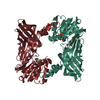
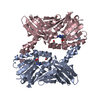
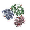
| Title | Crystal structure of human CARM1 with (R)-SKI-72 | ||||||
 Components Components | Histone-arginine methyltransferase CARM1 | ||||||
 Keywords Keywords | TRANSFERASE/TRANSFERASE INHIBITOR /  CARM1 / PRMT4 / SKI-72 inhibitor / Structural Genomics / Structural Genomics Consortium / SGC / TRANSFERASE-TRANSFERASE INHIBITOR complex CARM1 / PRMT4 / SKI-72 inhibitor / Structural Genomics / Structural Genomics Consortium / SGC / TRANSFERASE-TRANSFERASE INHIBITOR complex | ||||||
| Function / homology |  Function and homology information Function and homology information: / histone H3R17 methyltransferase activity / histone H3R2 methyltransferase activity / negative regulation of dendrite development / type I protein arginine methyltransferase / protein-arginine omega-N asymmetric methyltransferase activity / protein methyltransferase activity / histone arginine N-methyltransferase activity / regulation of intracellular estrogen receptor signaling pathway / replication fork reversal ...: / histone H3R17 methyltransferase activity / histone H3R2 methyltransferase activity / negative regulation of dendrite development / type I protein arginine methyltransferase / protein-arginine omega-N asymmetric methyltransferase activity / protein methyltransferase activity / histone arginine N-methyltransferase activity / regulation of intracellular estrogen receptor signaling pathway / replication fork reversal / protein-arginine N-methyltransferase activity / positive regulation of epithelial cell apoptotic process / histone methyltransferase activity / positive regulation of transcription by RNA polymerase I / nuclear replication fork / positive regulation of fat cell differentiation / response to cAMP / RORA activates gene expression / Regulation of lipid metabolism by PPARalpha / TP53 Regulates Transcription of Genes Involved in G2 Cell Cycle Arrest / BMAL1:CLOCK,NPAS2 activates circadian gene expression / Activation of gene expression by SREBF (SREBP) / nuclear receptor coactivator activity / lysine-acetylated histone binding / Heme signaling / Transcriptional activation of mitochondrial biogenesis / PPARA activates gene expression / Cytoprotection by HMOX1 / beta-catenin binding / RMTs methylate histone arginines / Transcriptional regulation of white adipocyte differentiation / Circadian Clock / DNA-binding transcription factor binding / Estrogen-dependent gene expression / transcription coactivator activity / transcription cis-regulatory region binding / positive regulation of cell population proliferation / regulation of DNA-templated transcription / nucleoplasm / nucleus / cytosol / cytoplasmSimilarity search - Function | ||||||
| Biological species |  Homo sapiens (human) Homo sapiens (human) | ||||||
| Method | X-RAY DIFFRACTION / SYNCHROTRON / MOLECULAR REPLACEMENT / molecular replacement / Resolution: 1.54 Å | ||||||
 Authors Authors | Dong, A. / Zeng, H. / Hutchinson, A. / Seitova, A. / Luo, M. / Cai, X.C. / Ke, W. / Wang, J. / Shi, C. / Zheng, W. ...Dong, A. / Zeng, H. / Hutchinson, A. / Seitova, A. / Luo, M. / Cai, X.C. / Ke, W. / Wang, J. / Shi, C. / Zheng, W. / Lee, J.P. / Ibanez, G. / Bountra, C. / Arrowsmith, C.H. / Edwards, A.M. / Brown, P.J. / Structural Genomics Consortium (SGC) | ||||||
 Citation Citation | Journal: to be published Title: Crystal structure of human CARM1 with (R)-SKI-72 Authors: Zeng, H. / Dong, A. / Hutchinson, A. / Seitova, A. / Luo, M. / Cai, X.C. / Ke, W. / Wang, J. / Shi, C. / Zheng, W. / Lee, J.P. / Ibanez, G. / Bountra, C. / Arrowsmith, C.H. / Edwards, A.M. / ...Authors: Zeng, H. / Dong, A. / Hutchinson, A. / Seitova, A. / Luo, M. / Cai, X.C. / Ke, W. / Wang, J. / Shi, C. / Zheng, W. / Lee, J.P. / Ibanez, G. / Bountra, C. / Arrowsmith, C.H. / Edwards, A.M. / Brown, P.J. / Structural Genomics Consortium (SGC) | ||||||
| History |
|
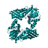
- Structure visualization
| Structure viewer | Molecule: MolmilJmol/JSmol |
|---|
- Downloads & links
-Download
| PDBx/mmCIF format | 6dvr.cif.gz | 162.8 KB | Display | PDBx/mmCIF format |
|---|---|---|---|---|
| PDB format | pdb6dvr.ent.gz | 126.1 KB | Display | PDB format |
| PDBx/mmJSON format | 6dvr.json.gz | Tree view | PDBx/mmJSON format | |
| Others |  Other downloads Other downloads |
-Validation report
| Arichive directory | https://data.pdbj.org/pub/pdb/validation_reports/dv/6dvrftp://data.pdbj.org/pub/pdb/validation_reports/dv/6dvr | HTTPS FTP |
|---|
-Related structure data
| Related structure data | 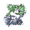 4ikpS S: Starting model for refinement |
|---|---|
| Similar structure data |
-Links
 PDBj
PDBj


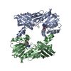
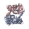
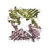
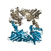
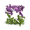
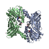
- Assembly
| Deposited unit | 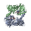
| ||||||||
|---|---|---|---|---|---|---|---|---|---|
| 1 |
| ||||||||
| 2 | 
| ||||||||
| Unit cell |
|
-Components
| #1: Protein | Mass: 38976.453 Da / Num. of mol.: 2 Source method: isolated from a genetically manipulated source Source: (gene. exp.) Homo sapiens (human) / Gene: CARM1, PRMT4 / Plasmid: pFBOH-MHL / Production host:   Spodoptera frugiperda (fall armyworm) / Strain (production host): Sf9 Spodoptera frugiperda (fall armyworm) / Strain (production host): Sf9References: UniProt: Q86X55, type I protein arginine methyltransferase #2: Chemical |   Mass: 618.726 Da / Num. of mol.: 2 / Source method: obtained synthetically / Formula: C32H42N8O5 Mass: 618.726 Da / Num. of mol.: 2 / Source method: obtained synthetically / Formula: C32H42N8O5#3: Chemical | 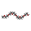  Mass: 296.357 Da / Num. of mol.: 2 / Source method: obtained synthetically / Formula: C13H28O7 Mass: 296.357 Da / Num. of mol.: 2 / Source method: obtained synthetically / Formula: C13H28O7#4: Chemical | ChemComp-UNX /   Num. of mol.: 6 / Source method: obtained synthetically Num. of mol.: 6 / Source method: obtained synthetically#5: Water | ChemComp-HOH / | Water Mass: 18.015 Da / Num. of mol.: 405 / Source method: isolated from a natural source / Formula: H2O Mass: 18.015 Da / Num. of mol.: 405 / Source method: isolated from a natural source / Formula: H2O |
|---|
-Experimental details
-Experiment
| Experiment | Method: X-RAY DIFFRACTION / Number of used crystals: 1 |
|---|
- Sample preparation
| Crystal | Density Matthews: 2.44 Å3/Da / Density % sol: 49.54 % / Mosaicity: 0.301 ° |
|---|---|
| Crystal grow | Temperature: 291 K / Method: vapor diffusion, hanging drop / pH: 7.5 / Details: 25% PEG 3350, 0.2 M NH4SO4, 0.1 M HEPES pH7.5 |
-Data collection
| Diffraction | Mean temperature: 100 K | |||||||||||||||||||||||||||||||||||||||||||||||||||||||||||||||||||||||||||||||||||||||||||||||||||||||||||||||||||||||||||||||||||||||||||||||||||||||||||||||||||||||||||||||||||||||||||||
|---|---|---|---|---|---|---|---|---|---|---|---|---|---|---|---|---|---|---|---|---|---|---|---|---|---|---|---|---|---|---|---|---|---|---|---|---|---|---|---|---|---|---|---|---|---|---|---|---|---|---|---|---|---|---|---|---|---|---|---|---|---|---|---|---|---|---|---|---|---|---|---|---|---|---|---|---|---|---|---|---|---|---|---|---|---|---|---|---|---|---|---|---|---|---|---|---|---|---|---|---|---|---|---|---|---|---|---|---|---|---|---|---|---|---|---|---|---|---|---|---|---|---|---|---|---|---|---|---|---|---|---|---|---|---|---|---|---|---|---|---|---|---|---|---|---|---|---|---|---|---|---|---|---|---|---|---|---|---|---|---|---|---|---|---|---|---|---|---|---|---|---|---|---|---|---|---|---|---|---|---|---|---|---|---|---|---|---|---|---|---|
| Diffraction source | Source: SYNCHROTRON / Site: APS  / Beamline: 24-ID-E / Wavelength: 0.9789 Å / Beamline: 24-ID-E / Wavelength: 0.9789 Å | |||||||||||||||||||||||||||||||||||||||||||||||||||||||||||||||||||||||||||||||||||||||||||||||||||||||||||||||||||||||||||||||||||||||||||||||||||||||||||||||||||||||||||||||||||||||||||||
| Detector | Type: DECTRIS EIGER X 16M / Detector: PIXEL / Date: Apr 8, 2018 | |||||||||||||||||||||||||||||||||||||||||||||||||||||||||||||||||||||||||||||||||||||||||||||||||||||||||||||||||||||||||||||||||||||||||||||||||||||||||||||||||||||||||||||||||||||||||||||
| Radiation | Protocol: SINGLE WAVELENGTH / Monochromatic (M) / Laue (L): M / Scattering type: x-ray | |||||||||||||||||||||||||||||||||||||||||||||||||||||||||||||||||||||||||||||||||||||||||||||||||||||||||||||||||||||||||||||||||||||||||||||||||||||||||||||||||||||||||||||||||||||||||||||
| Radiation wavelength | Wavelength: 0.9789 Å / Relative weight: 1 | |||||||||||||||||||||||||||||||||||||||||||||||||||||||||||||||||||||||||||||||||||||||||||||||||||||||||||||||||||||||||||||||||||||||||||||||||||||||||||||||||||||||||||||||||||||||||||||
| Reflection | Resolution: 1.54→50 Å / Num. obs: 112857 / % possible obs: 100 % / Redundancy: 10.7 % / Rmerge(I) obs: 0.049 / Rpim(I) all: 0.016 / Rrim(I) all: 0.052 / Χ2: 0.624 / Net I/σ(I): 7.3 | |||||||||||||||||||||||||||||||||||||||||||||||||||||||||||||||||||||||||||||||||||||||||||||||||||||||||||||||||||||||||||||||||||||||||||||||||||||||||||||||||||||||||||||||||||||||||||||
| Reflection shell | Diffraction-ID: 1
|
-Phasing
| Phasing | Method: molecular replacement |
|---|
- Processing
| Software |
| ||||||||||||||||||||||||||||||||||||||||||||||||||||||||||||
|---|---|---|---|---|---|---|---|---|---|---|---|---|---|---|---|---|---|---|---|---|---|---|---|---|---|---|---|---|---|---|---|---|---|---|---|---|---|---|---|---|---|---|---|---|---|---|---|---|---|---|---|---|---|---|---|---|---|---|---|---|---|
| Refinement | Method to determine structure: MOLECULAR REPLACEMENT Starting model: 4IKP Resolution: 1.54→44.56 Å / Cor.coef. Fo:Fc: 0.964 / Cor.coef. Fo:Fc free: 0.957 / SU B: 1.572 / SU ML: 0.057 / Cross valid method: THROUGHOUT / σ(F): 0 / ESU R: 0.08 / ESU R Free: 0.078 Details: HYDROGENS HAVE BEEN ADDED IN THE RIDING POSITIONS U VALUES : REFINED INDIVIDUALLY
| ||||||||||||||||||||||||||||||||||||||||||||||||||||||||||||
| Solvent computation | Ion probe radii: 0.8 Å / Shrinkage radii: 0.8 Å / VDW probe radii: 1.2 Å | ||||||||||||||||||||||||||||||||||||||||||||||||||||||||||||
| Displacement parameters | Biso max: 61.9 Å2 / Biso mean: 23.388 Å2 / Biso min: 12.3 Å2
| ||||||||||||||||||||||||||||||||||||||||||||||||||||||||||||
| Refinement step | Cycle: final / Resolution: 1.54→44.56 Å
| ||||||||||||||||||||||||||||||||||||||||||||||||||||||||||||
| Refine LS restraints |
| ||||||||||||||||||||||||||||||||||||||||||||||||||||||||||||
| LS refinement shell | Resolution: 1.54→1.58 Å / Rfactor Rfree error: 0 / Total num. of bins used: 20
|
