 Movie
Movie Controller


+ Open data

- Basic information
| Entry | Database: PDB / ID: 6dtu | |||||||||
|---|---|---|---|---|---|---|---|---|---|---|
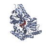
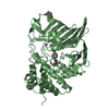
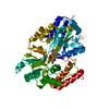
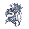
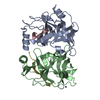
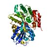
| Title | Maltotetraose bound T. maritima MalE1 | |||||||||
 Components Components | maltose-binding protein MalE1 | |||||||||
 Keywords Keywords | SUGAR BINDING PROTEIN / periplasmic binding protein / maltose binding protein / maltotetraose | |||||||||
| Function / homology |  Function and homology information Function and homology informationcarbohydrate transmembrane transporter activity / maltose binding / maltose transport / maltodextrin transmembrane transport / proteasome core complex, alpha-subunit complex / ATP-binding cassette (ABC) transporter complex, substrate-binding subunit-containing / ubiquitin-dependent protein catabolic process Similarity search - Function | |||||||||
| Biological species |   Thermotoga maritima (bacteria) Thermotoga maritima (bacteria) | |||||||||
| Method |  X-RAY DIFFRACTION / MOLECULAR REPLACEMENT / Resolution: 1.5 Å X-RAY DIFFRACTION / MOLECULAR REPLACEMENT / Resolution: 1.5 Å | |||||||||
 Authors Authors | Cuneo, M.J. / Shukla, S. | |||||||||
 Citation Citation | Journal: Biochemistry / Year: 2018 Title: Differential Substrate Recognition by Maltose Binding Proteins Influenced by Structure and Dynamics. Authors: Shukla, S. / Bafna, K. / Gullett, C. / Myles, D.A.A. / Agarwal, P.K. / Cuneo, M.J. | |||||||||
| History |
|
- Structure visualization
| Structure viewer | Molecule: MolmilJmol/JSmol |
|---|
- Downloads & links
-Download
| PDBx/mmCIF format | 6dtu.cif.gz | 172.3 KB | Display | PDBx/mmCIF format |
|---|---|---|---|---|
| PDB format | pdb6dtu.ent.gz | 134.9 KB | Display | PDB format |
| PDBx/mmJSON format | 6dtu.json.gz | Tree view | PDBx/mmJSON format | |
| Others |  Other downloads Other downloads |
-Validation report
| Summary document | 6dtu_validation.pdf.gz | 1.1 MB | Display | wwPDB validaton report |
|---|---|---|---|---|
| Full document | 6dtu_full_validation.pdf.gz | 1.1 MB | Display | |
| Data in XML | 6dtu_validation.xml.gz | 32.3 KB | Display | |
| Data in CIF | 6dtu_validation.cif.gz | 48.7 KB | Display | |
| Arichive directory | https://data.pdbj.org/pub/pdb/validation_reports/dt/6dtuftp://data.pdbj.org/pub/pdb/validation_reports/dt/6dtu | HTTPS FTP |
-Related structure data
| Related structure data |  6dtqC 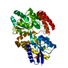 6dtrC  6dtsC  6dttC  2ghaS S: Starting model for refinement C: citing same article ( |
|---|---|
| Similar structure data |


-Links
 PDBj
PDBj


- Assembly
| Deposited unit | 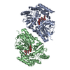
| ||||||||
|---|---|---|---|---|---|---|---|---|---|
| 1 | 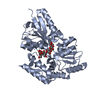
| ||||||||
| 2 | 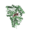
| ||||||||
| Unit cell |
|
-Components
| #1: Protein | Mass: 41334.316 Da / Num. of mol.: 2 Source method: isolated from a genetically manipulated source Source: (gene. exp.) Thermotoga maritima (strain ATCC 43589 / MSB8 / DSM 3109 / JCM 10099) (bacteria)Strain: ATCC 43589 / MSB8 / DSM 3109 / JCM 10099 / Gene: TM_1204 / Production host: #2: Polysaccharide | 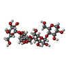  Source method: isolated from a genetically manipulated source Details: oligosaccharide / References: alpha-maltotetraose #3: Water | ChemComp-HOH / |  Mass: 18.015 Da / Num. of mol.: 524 / Source method: isolated from a natural source / Formula: H2O Mass: 18.015 Da / Num. of mol.: 524 / Source method: isolated from a natural source / Formula: H2O |
|---|
-Experimental details
-Experiment
| Experiment | Method: X-RAY DIFFRACTION / Number of used crystals: 1 |
|---|
- Sample preparation
| Crystal | Density Matthews: 2.1 Å3/Da / Density % sol: 41.52 % |
|---|---|
| Crystal grow | Temperature: 293 K / Method: vapor diffusion, hanging drop / pH: 7.5 Details: 25% poly-ethylene glycol (PEG) 4000, 18% isopropanol and 0.1M sodium citrate |
-Data collection
| Diffraction | Mean temperature: 100 K |
|---|---|
| Diffraction source | Source: ROTATING ANODE / Type: RIGAKU MICROMAX-007 HF / Wavelength: 1.5418 Å |
| Detector | Type: RIGAKU RAXIS IV++ / Detector: IMAGE PLATE / Date: Jun 12, 2018 |
| Radiation | Protocol: SINGLE WAVELENGTH / Monochromatic (M) / Laue (L): M / Scattering type: x-ray |
| Radiation wavelength | Wavelength: 1.5418 Å / Relative weight: 1 |
| Reflection | Resolution: 1.5→50 Å / Num. obs: 90224 / % possible obs: 83.4 % / Redundancy: 2.1 % / Rmerge(I) obs: 0.077 / Net I/σ(I): 3 |
| Reflection shell | Resolution: 1.5→1.55 Å / Rmerge(I) obs: 0.464 / Num. unique obs: 8982 |
- Processing
| Software |
| |||||||||||||||||||||||||||||||||||||||||||||||||||||||||||||||||||||||||||||||||||||||||||||||||||||||||
|---|---|---|---|---|---|---|---|---|---|---|---|---|---|---|---|---|---|---|---|---|---|---|---|---|---|---|---|---|---|---|---|---|---|---|---|---|---|---|---|---|---|---|---|---|---|---|---|---|---|---|---|---|---|---|---|---|---|---|---|---|---|---|---|---|---|---|---|---|---|---|---|---|---|---|---|---|---|---|---|---|---|---|---|---|---|---|---|---|---|---|---|---|---|---|---|---|---|---|---|---|---|---|---|---|---|---|
| Refinement | Method to determine structure: MOLECULAR REPLACEMENT Starting model: 2GHA Resolution: 1.5→30.816 Å / SU ML: 0.19 / Cross valid method: FREE R-VALUE / σ(F): 1.96 / Phase error: 23.73
| |||||||||||||||||||||||||||||||||||||||||||||||||||||||||||||||||||||||||||||||||||||||||||||||||||||||||
| Solvent computation | Shrinkage radii: 0.9 Å / VDW probe radii: 1.11 Å | |||||||||||||||||||||||||||||||||||||||||||||||||||||||||||||||||||||||||||||||||||||||||||||||||||||||||
| Refinement step | Cycle: LAST / Resolution: 1.5→30.816 Å
| |||||||||||||||||||||||||||||||||||||||||||||||||||||||||||||||||||||||||||||||||||||||||||||||||||||||||
| Refine LS restraints |
| |||||||||||||||||||||||||||||||||||||||||||||||||||||||||||||||||||||||||||||||||||||||||||||||||||||||||
| LS refinement shell |
|
