 Movie
Movie Controller

[English] 日本語
 Yorodumi
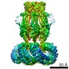
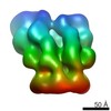
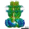
- PDB-6djb: Structure of human Volume Regulated Anion Channel composed of SWE... -
+ Open data

- Basic information
| Entry | Database: PDB / ID: 6djb | ||||||||||||
|---|---|---|---|---|---|---|---|---|---|---|---|---|---|
| Title | Structure of human Volume Regulated Anion Channel composed of SWELL1 (LRRC8A) | ||||||||||||
 Components Components | Volume-regulated anion channel subunit LRRC8A | ||||||||||||
 Keywords Keywords | MEMBRANE PROTEIN / anion channel / LRRC8A / SWELL1 | ||||||||||||
| Function / homology |  Function and homology information Function and homology informationMiscellaneous transport and binding events / pre-B cell differentiation / volume-sensitive anion channel activity / taurine transmembrane transport / monoatomic anion transmembrane transport / aspartate transmembrane transport / cyclic-GMP-AMP transmembrane transporter activity / cyclic-GMP-AMP transmembrane import across plasma membrane / monoatomic anion transport / cell volume homeostasis ...Miscellaneous transport and binding events / pre-B cell differentiation / volume-sensitive anion channel activity / taurine transmembrane transport / monoatomic anion transmembrane transport / aspartate transmembrane transport / cyclic-GMP-AMP transmembrane transporter activity / cyclic-GMP-AMP transmembrane import across plasma membrane / monoatomic anion transport / cell volume homeostasis / protein hexamerization / response to osmotic stress / monoatomic ion channel complex / intracellular glucose homeostasis / positive regulation of myoblast differentiation / chloride transmembrane transport / positive regulation of insulin secretion / spermatogenesis / lysosomal membrane / cell surface / signal transduction / membrane / identical protein binding / plasma membraneSimilarity search - Function | ||||||||||||
| Biological species |  Homo sapiens (human) Homo sapiens (human) | ||||||||||||
| Method | ELECTRON MICROSCOPY / single particle reconstruction / cryo EM / Resolution: 4.4 Å | ||||||||||||
 Authors Authors | Kefauver, J.M. / Saotome, K. / Pallesen, J. / Cottrell, C.A. / Ward, A.B. / Patapoutian, A. | ||||||||||||
| Funding support |  United States, 3items United States, 3items
| ||||||||||||
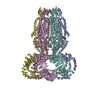
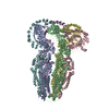
 Citation Citation | Journal: Elife / Year: 2018 Title: Structure of the human volume regulated anion channel. Authors: Jennifer M Kefauver / Kei Saotome / Adrienne E Dubin / Jesper Pallesen / Christopher A Cottrell / Stuart M Cahalan / Zhaozhu Qiu / Gunhee Hong / Christopher S Crowley / Tess Whitwam / Wen- ...Authors: Jennifer M Kefauver / Kei Saotome / Adrienne E Dubin / Jesper Pallesen / Christopher A Cottrell / Stuart M Cahalan / Zhaozhu Qiu / Gunhee Hong / Christopher S Crowley / Tess Whitwam / Wen-Hsin Lee / Andrew B Ward / Ardem Patapoutian / Abstract: SWELL1 (LRRC8A) is the only essential subunit of the Volume Regulated Anion Channel (VRAC), which regulates cellular volume homeostasis and is activated by hypotonic solutions. SWELL1, together with ...SWELL1 (LRRC8A) is the only essential subunit of the Volume Regulated Anion Channel (VRAC), which regulates cellular volume homeostasis and is activated by hypotonic solutions. SWELL1, together with four other LRRC8 family members, potentially forms a vastly heterogeneous cohort of VRAC channels with different properties; however, SWELL1 alone is also functional. Here, we report a high-resolution cryo-electron microscopy structure of full-length human homo-hexameric SWELL1. The structure reveals a trimer of dimers assembly with symmetry mismatch between the pore-forming domain and the cytosolic leucine-rich repeat (LRR) domains. Importantly, mutational analysis demonstrates that a charged residue at the narrowest constriction of the homomeric channel is an important pore determinant of heteromeric VRAC. Additionally, a mutation in the flexible N-terminal portion of SWELL1 affects pore properties, suggesting a putative link between intracellular structures and channel regulation. This structure provides a scaffold for further dissecting the heterogeneity and mechanism of activation of VRAC. | ||||||||||||
| History |
|
- Structure visualization
| Movie |
Movie viewer |
|---|---|
| Structure viewer | Molecule: MolmilJmol/JSmol |
- Downloads & links
-Download
| PDBx/mmCIF format | 6djb.cif.gz | 832.9 KB | Display | PDBx/mmCIF format |
|---|---|---|---|---|
| PDB format | pdb6djb.ent.gz | 703 KB | Display | PDB format |
| PDBx/mmJSON format | 6djb.json.gz | Tree view | PDBx/mmJSON format | |
| Others |  Other downloads Other downloads |
-Validation report
| Arichive directory | https://data.pdbj.org/pub/pdb/validation_reports/dj/6djbftp://data.pdbj.org/pub/pdb/validation_reports/dj/6djb | HTTPS FTP |
|---|
-Related structure data
| Related structure data | 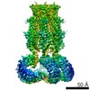 7935MC M: map data used to model this data C: citing same article ( |
|---|---|
| Similar structure data |


-Links
 PDBj
PDBj


- Assembly
| Deposited unit | 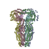
|
|---|---|
| 1 |
|
-Components
| #1: Protein | / Leucine-rich repeat-containing protein 8A / Swelling protein 1 Mass: 94318.516 Da / Num. of mol.: 6 Source method: isolated from a genetically manipulated source Source: (gene. exp.) Homo sapiens (human) / Gene: LRRC8A, KIAA1437, LRRC8, SWELL1, UNQ221/PRO247 / Cell line (production host): HEK 293F LRRC8(B,C,D,E)-/- / Production host: Homo sapiens (human) / References: UniProt: Q8IWT6 |
|---|
-Experimental details
-Experiment
| Experiment | Method: ELECTRON MICROSCOPY |
|---|---|
| EM experiment | Aggregation state: PARTICLE / 3D reconstruction method: single particle reconstruction |
- Sample preparation
| Component | Name: Homomeric Volume Regulated Anion Channel composed of SWELL1 Type: COMPLEX / Entity ID: all / Source: RECOMBINANT | |||||||||||||||||||||||||
|---|---|---|---|---|---|---|---|---|---|---|---|---|---|---|---|---|---|---|---|---|---|---|---|---|---|---|
| Molecular weight | Experimental value: NO | |||||||||||||||||||||||||
| Source (natural) | Organism: Homo sapiens (human) | |||||||||||||||||||||||||
| Source (recombinant) | Organism: Homo sapiens (human) / Cell: HEK 293F LRRC8(B,C,D,E)-/- | |||||||||||||||||||||||||
| Buffer solution | pH: 8 | |||||||||||||||||||||||||
| Buffer component |
| |||||||||||||||||||||||||
| Specimen | Conc.: 8 mg/ml / Embedding applied: NO / Shadowing applied: NO / Staining applied: NO / Vitrification applied: YES | |||||||||||||||||||||||||
| Specimen support | Grid material: GOLD / Grid type: Quantifoil, UltrAuFoil, R1.2/1.3 | |||||||||||||||||||||||||
| Vitrification | Instrument: FEI VITROBOT MARK IV / Cryogen name: ETHANE / Humidity: 100 % / Chamber temperature: 277 K |
- Electron microscopy imaging
| Experimental equipment |  Model: Talos Arctica / Image courtesy: FEI Company |
|---|---|
| Microscopy | Model: FEI TALOS ARCTICA |
| Electron gun | Electron source: FIELD EMISSION GUN / Accelerating voltage: 200 kV / Illumination mode: FLOOD BEAM |
| Electron lens | Mode: BRIGHT FIELDBright-field microscopy / Nominal magnification: 36000 X / Nominal defocus max: 1500 nm / Nominal defocus min: 800 nm / Cs: 2.7 mm / C2 aperture diameter: 70 µm |
| Specimen holder | Cryogen: NITROGEN |
| Image recording | Electron dose: 55 e/Å2 / Detector mode: COUNTING / Film or detector model: GATAN K2 SUMMIT (4k x 4k) / Num. of grids imaged: 1 / Num. of real images: 4627 |
| Image scans | Width: 3710 / Height: 3838 |
- Processing
| EM software |
| ||||||||||||
|---|---|---|---|---|---|---|---|---|---|---|---|---|---|
| CTF correction | Type: PHASE FLIPPING AND AMPLITUDE CORRECTION | ||||||||||||
| Particle selection | Num. of particles selected: 316206 | ||||||||||||
| Symmetry | Point symmetry: C3 (3 fold cyclic) | ||||||||||||
| 3D reconstruction | Resolution: 4.4 Å / Resolution method: FSC 0.143 CUT-OFF / Num. of particles: 25719 / Symmetry type: POINT |
