 Movie
Movie Controller


+ Open data

- Basic information
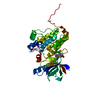
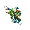
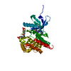
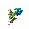
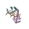
| Entry | Database: PDB / ID: 6dei | ||||||
|---|---|---|---|---|---|---|---|
| Title | Structure of Dse3-Csm1 complex | ||||||
 Components Components |
| ||||||
 Keywords Keywords | Protein Binding/Cell Cycle /  monopolin / daughter cell-specific protein / Protein Binding-Cell Cycle complex monopolin / daughter cell-specific protein / Protein Binding-Cell Cycle complex | ||||||
| Function / homology |  Function and homology informationmonopolin complex / spindle attachment to meiosis I kinetochore / protein localization to nucleolar rDNA repeats / meiotic chromosome segregation / meiotic sister chromatid cohesion, centromeric / rDNA chromatin condensation / homologous chromosome segregation / cellular bud neck / nuclear envelope / cell cycle ...monopolin complex / spindle attachment to meiosis I kinetochore / protein localization to nucleolar rDNA repeats / meiotic chromosome segregation / meiotic sister chromatid cohesion, centromeric / rDNA chromatin condensation / homologous chromosome segregation / cellular bud neck / nuclear envelope / cell cycle / nucleolus / identical protein binding / nucleus / cytoplasm Function and homology informationmonopolin complex / spindle attachment to meiosis I kinetochore / protein localization to nucleolar rDNA repeats / meiotic chromosome segregation / meiotic sister chromatid cohesion, centromeric / rDNA chromatin condensation / homologous chromosome segregation / cellular bud neck / nuclear envelope / cell cycle ...monopolin complex / spindle attachment to meiosis I kinetochore / protein localization to nucleolar rDNA repeats / meiotic chromosome segregation / meiotic sister chromatid cohesion, centromeric / rDNA chromatin condensation / homologous chromosome segregation / cellular bud neck / nuclear envelope / cell cycle / nucleolus / identical protein binding / nucleus / cytoplasmSimilarity search - Function | ||||||
| Biological species |  Saccharomyces cerevisiae (brewer's yeast) Saccharomyces cerevisiae (brewer's yeast) | ||||||
| Method | X-RAY DIFFRACTION / SYNCHROTRON / MOLECULAR REPLACEMENT / Resolution: 1.699 Å | ||||||
 Authors Authors | Singh, N. / Corbett, K.D. | ||||||
| Funding support |  United States, 1items United States, 1items
| ||||||
 Citation Citation | Journal: Protein Sci. / Year: 2018 Title: The budding-yeast RWD protein Csm1 scaffolds diverse protein complexes through a conserved structural mechanism. Authors: Singh, N. / Corbett, K.D. | ||||||
| History |
|
- Structure visualization
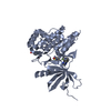
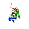
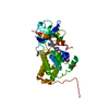
| Structure viewer | Molecule: MolmilJmol/JSmol |
|---|
- Downloads & links
-Download
| PDBx/mmCIF format | 6dei.cif.gz | 172.4 KB | Display | PDBx/mmCIF format |
|---|---|---|---|---|
| PDB format | pdb6dei.ent.gz | 137.6 KB | Display | PDB format |
| PDBx/mmJSON format | 6dei.json.gz | Tree view | PDBx/mmJSON format | |
| Others |  Other downloads Other downloads |
-Validation report
| Arichive directory | https://data.pdbj.org/pub/pdb/validation_reports/de/6deiftp://data.pdbj.org/pub/pdb/validation_reports/de/6dei | HTTPS FTP |
|---|
-Related structure data
| Related structure data | 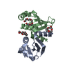 3n4sS S: Starting model for refinement |
|---|---|
| Similar structure data | |
| Experimental dataset #1 | Data reference: 10.15785/SBGRID/585 / Data set type: diffraction image data |

-Links
 PDBj
PDBj- Assembly
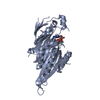
| Deposited unit | 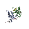
| ||||||||
|---|---|---|---|---|---|---|---|---|---|
| 1 |
| ||||||||
| Unit cell |
|
-Components
| #1: Protein | / Chromosome segregation in meiosis protein 1 Mass: 12991.447 Da / Num. of mol.: 2 Source method: isolated from a genetically manipulated source Source: (gene. exp.) Saccharomyces cerevisiae (strain ATCC 204508 / S288c) (yeast)Strain: ATCC 204508 / S288c / Gene: CSM1, SPO86, YCR086W, YCR86W / Production host:  Escherichia coli (E. coli) / References: UniProt: P25651 Escherichia coli (E. coli) / References: UniProt: P25651#2: Protein/peptide | Mass: 2705.136 Da / Num. of mol.: 2 Source method: isolated from a genetically manipulated source Source: (gene. exp.) Saccharomyces cerevisiae (strain ATCC 204508 / S288c) (yeast)Strain: ATCC 204508 / S288c / Gene: DSE3, YOR264W / Production host: Escherichia coli (E. coli) / References: UniProt: Q08729#3: Chemical | Polyethylene glycol  Mass: 150.173 Da / Num. of mol.: 2 / Source method: obtained synthetically / Formula: C6H14O4 Mass: 150.173 Da / Num. of mol.: 2 / Source method: obtained synthetically / Formula: C6H14O4#4: Chemical | ChemComp-ACT / | Acetate  Mass: 59.044 Da / Num. of mol.: 1 / Source method: obtained synthetically / Formula: C2H3O2 Mass: 59.044 Da / Num. of mol.: 1 / Source method: obtained synthetically / Formula: C2H3O2#5: Water | ChemComp-HOH / | Water Mass: 18.015 Da / Num. of mol.: 232 / Source method: isolated from a natural source / Formula: H2O Mass: 18.015 Da / Num. of mol.: 232 / Source method: isolated from a natural source / Formula: H2O |
|---|
-Experimental details
-Experiment
| Experiment | Method: X-RAY DIFFRACTION / Number of used crystals: 1 |
|---|
- Sample preparation
| Crystal | Density Matthews: 1.99 Å3/Da / Density % sol: 38.27 % |
|---|---|
| Crystal grow | Temperature: 293 K / Method: vapor diffusion, hanging drop / Details: 0.1M NaOAc, 0.1M HEPES pH 7.5, 22% PEG 4000 |
-Data collection
| Diffraction | Mean temperature: 100 K |
|---|---|
| Diffraction source | Source: SYNCHROTRON / Site: SSRL / Beamline: BL9-2 / Wavelength: 0.99833 Å |
| Detector | Type: DECTRIS PILATUS 6M / Detector: PIXEL / Date: Feb 19, 2017 |
| Radiation | Protocol: SINGLE WAVELENGTH / Monochromatic (M) / Laue (L): M / Scattering type: x-ray |
| Radiation wavelength | Wavelength: 0.99833 Å / Relative weight: 1 |
| Reflection | Resolution: 1.699→39.3 Å / Num. obs: 28421 / % possible obs: 99.1 % / Redundancy: 6.5 % / CC1/2: 0.997 / Net I/σ(I): 8.3 |
| Reflection shell | Resolution: 1.699→1.73 Å / Num. unique all: 1451 / CC1/2: 0.436 / % possible all: 95.1 |
- Processing
| Software |
| |||||||||||||||||||||||||||||||||||||||||||||||||||||||||||||||||||||||||||||||||||||||||||||||||||||||||
|---|---|---|---|---|---|---|---|---|---|---|---|---|---|---|---|---|---|---|---|---|---|---|---|---|---|---|---|---|---|---|---|---|---|---|---|---|---|---|---|---|---|---|---|---|---|---|---|---|---|---|---|---|---|---|---|---|---|---|---|---|---|---|---|---|---|---|---|---|---|---|---|---|---|---|---|---|---|---|---|---|---|---|---|---|---|---|---|---|---|---|---|---|---|---|---|---|---|---|---|---|---|---|---|---|---|---|
| Refinement | Method to determine structure: MOLECULAR REPLACEMENT Starting model: 3N4S Resolution: 1.699→39.095 Å / SU ML: 0.23 / Cross valid method: FREE R-VALUE / σ(F): 1.33 / Phase error: 22.04 / Stereochemistry target values: ML
| |||||||||||||||||||||||||||||||||||||||||||||||||||||||||||||||||||||||||||||||||||||||||||||||||||||||||
| Solvent computation | Shrinkage radii: 0.9 Å / VDW probe radii: 1.11 Å / Solvent model: FLAT BULK SOLVENT MODEL | |||||||||||||||||||||||||||||||||||||||||||||||||||||||||||||||||||||||||||||||||||||||||||||||||||||||||
| Refinement step | Cycle: LAST / Resolution: 1.699→39.095 Å
| |||||||||||||||||||||||||||||||||||||||||||||||||||||||||||||||||||||||||||||||||||||||||||||||||||||||||
| Refine LS restraints |
| |||||||||||||||||||||||||||||||||||||||||||||||||||||||||||||||||||||||||||||||||||||||||||||||||||||||||
| LS refinement shell |
| |||||||||||||||||||||||||||||||||||||||||||||||||||||||||||||||||||||||||||||||||||||||||||||||||||||||||
| Refinement TLS params. | Method: refined / Origin x: -7.7943 Å / Origin y: 1.2655 Å / Origin z: 11.6193 Å
| |||||||||||||||||||||||||||||||||||||||||||||||||||||||||||||||||||||||||||||||||||||||||||||||||||||||||
| Refinement TLS group | Selection details: all |
