[English] 日本語
Yorodumi
- PDB-6d97: Structure of aldehyde dehydrogenase 12 (ALDH12) from Zea mays -

+
Open data


ID or keywords:

Loading...

-
Basic information

Entry
Database: PDB / ID: 6d97
TitleStructure of aldehyde dehydrogenase 12 (ALDH12) from Zea mays
ComponentsAldehyde dehydrogenase 12
KeywordsOXIDOREDUCTASE / ALDH12 / ROSSMANN FOLD / L-glutamate-gamma-semialdehyde dehydrogenase
Function / homology
Function and homology information


cellular response to chemical stimulus / 1-pyrroline-5-carboxylate dehydrogenase activity / L-glutamate gamma-semialdehyde dehydrogenase / aldehyde dehydrogenase (NAD+) activity
Similarity search - Function
Aldehyde dehydrogenase family 7 member A1-like / Aldehyde dehydrogenase, cysteine active site / Aldehyde dehydrogenases cysteine active site. / Aldehyde dehydrogenase domain / Aldehyde dehydrogenase family / Aldehyde dehydrogenase, N-terminal / Aldehyde dehydrogenase, C-terminal / Aldehyde/histidinol dehydrogenase
Similarity search - Domain/homology
NICOTINAMIDE-ADENINE-DINUCLEOTIDE / Aldehyde dehydrogenase 12
Similarity search - Component
Biological speciesZea mays (maize)
MethodX-RAY DIFFRACTION / SYNCHROTRON / MOLECULAR REPLACEMENT / Resolution: 2.2 Å
AuthorsTanner, J.J. / Korasick, D.A. / Kopecny, D.
Funding support United States, 2items
OrganizationGrant numberCountry
National Institutes of Health/National Institute of General Medical Sciences (NIH/NIGMS)R01GM065546 United States
National Institutes of Health/National Institute of General Medical Sciences (NIH/NIGMS)R01GM093123 United States
Citation
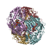
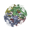
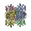
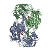
Journal: J Mol Biol / Year: 2019
Title: Structural and Biochemical Characterization of Aldehyde Dehydrogenase 12, the Last Enzyme of Proline Catabolism in Plants.
Authors: David A Korasick / Radka Končitíková / Martina Kopečná / Eva Hájková / Armelle Vigouroux / Solange Moréra / Donald F Becker / Marek Šebela / John J Tanner / David Kopečný /
Abstract: Heterokonts, Alveolata protists, green algae from Charophyta and Chlorophyta divisions, and all Embryophyta plants possess an aldehyde dehydrogenase (ALDH) gene named ALDH12. Here, we provide a ...Heterokonts, Alveolata protists, green algae from Charophyta and Chlorophyta divisions, and all Embryophyta plants possess an aldehyde dehydrogenase (ALDH) gene named ALDH12. Here, we provide a biochemical characterization of two ALDH12 family members from the lower plant Physcomitrella patens and higher plant Zea mays. We show that ALDH12 encodes an NAD-dependent glutamate γ-semialdehyde dehydrogenase (GSALDH), which irreversibly converts glutamate γ-semialdehyde (GSAL), a mitochondrial intermediate of the proline and arginine catabolism, to glutamate. Sedimentation equilibrium and small-angle X-ray scattering analyses reveal that in solution both plant GSALDHs exist as equilibrium between a domain-swapped dimer and the dimer-of-dimers tetramer. Plant GSALDHs share very low-sequence identity with bacterial, fungal, and animal GSALDHs (classified as ALDH4), which are the closest related ALDH superfamily members. Nevertheless, the crystal structure of ZmALDH12 at 2.2-Å resolution  shows that nearly all key residues involved in the recognition of GSAL are identical to those in ALDH4, indicating a close functional relationship with ALDH4. Phylogenetic analysis suggests that the transition from ALDH4 to ALDH12 occurred during the evolution of the endosymbiotic plant ancestor, prior to the evolution of green algae and land plants. Finally, ALDH12 expression in maize and moss is downregulated in response to salt and drought stresses, possibly to maintain proline levels. Taken together, these results provide molecular insight into the biological roles of the plant ALDH12 family.
History
DepositionApr 27, 2018Deposition site: RCSB / Processing site: RCSB
Revision 1.0Jan 9, 2019Provider: repository / Type: Initial release
Revision 1.1Feb 6, 2019Group: Data collection / Database references / Category: citation
Item: _citation.journal_volume / _citation.page_first ..._citation.journal_volume / _citation.page_first / _citation.page_last / _citation.year
Revision 1.2Jan 1, 2020Group: Author supporting evidence / Category: pdbx_audit_support / Item: _pdbx_audit_support.funding_organization
Revision 1.3Oct 4, 2023Group: Data collection / Database references / Refinement description
Category: chem_comp_atom / chem_comp_bond ...chem_comp_atom / chem_comp_bond / database_2 / pdbx_initial_refinement_model
Item: _database_2.pdbx_DOI / _database_2.pdbx_database_accession

-
Structure visualization

Structure viewerMolecule:
MolmilJmol/JSmol

Downloads & links

-
Assembly

Deposited unit
A: Aldehyde dehydrogenase 12
B: Aldehyde dehydrogenase 12
C: Aldehyde dehydrogenase 12
D: Aldehyde dehydrogenase 12
hetero molecules


Theoretical massNumber of molelcules
Total (without water)244,5188
Polymers241,8654
Non-polymers2,6544
Water10,413578
1
A: Aldehyde dehydrogenase 12
B: Aldehyde dehydrogenase 12
hetero molecules

A: Aldehyde dehydrogenase 12
B: Aldehyde dehydrogenase 12
hetero molecules


Theoretical massNumber of molelcules
Total (without water)244,5188
Polymers241,8654
Non-polymers2,6544
Water724
TypeNameSymmetry operationNumber
identity operation1_555x,y,z1
crystal symmetry operation2_556-x,y,-z+11
Buried area26300 Å2
ΔGint-70 kcal/mol
Surface area60680 Å2
MethodPISA
2
A: Aldehyde dehydrogenase 12
B: Aldehyde dehydrogenase 12
hetero molecules


Theoretical massNumber of molelcules
Total (without water)122,2594
Polymers120,9322
Non-polymers1,3272
Water362
TypeNameSymmetry operationNumber
identity operation1_555x,y,z1
Buried area8900 Å2
ΔGint-23 kcal/mol
Surface area34590 Å2
MethodPISA
3
C: Aldehyde dehydrogenase 12
D: Aldehyde dehydrogenase 12
hetero molecules

C: Aldehyde dehydrogenase 12
D: Aldehyde dehydrogenase 12
hetero molecules


Theoretical massNumber of molelcules
Total (without water)244,5188
Polymers241,8654
Non-polymers2,6544
Water724
TypeNameSymmetry operationNumber
identity operation1_555x,y,z1
crystal symmetry operation2_657-x+1,y,-z+21
Buried area26240 Å2
ΔGint-66 kcal/mol
Surface area61110 Å2
MethodPISA
4
C: Aldehyde dehydrogenase 12
D: Aldehyde dehydrogenase 12
hetero molecules


Theoretical massNumber of molelcules
Total (without water)122,2594
Polymers120,9322
Non-polymers1,3272
Water362
TypeNameSymmetry operationNumber
identity operation1_555x,y,z1
Buried area8960 Å2
ΔGint-22 kcal/mol
Surface area34710 Å2
MethodPISA
Unit cell
Length a, b, c (Å)160.642, 123.737, 103.707
Angle α, β, γ (deg.)90.000, 105.620, 90.000
Int Tables number5
Space group name H-MC121

-
Components

#1: Protein
Aldehyde dehydrogenase 12 /


Mass: 60466.184 Da / Num. of mol.: 4
Source method: isolated from a genetically manipulated source
Source: (gene. exp.) Zea mays (maize) / Gene: ALDH12 / Production host: Escherichia coli (E. coli)
References: UniProt: A0A2H4PMI3, L-glutamate gamma-semialdehyde dehydrogenase
#2: Chemical
ChemComp-NAD / NICOTINAMIDE-ADENINE-DINUCLEOTIDE / Nicotinamide adenine dinucleotide


Mass: 663.425 Da / Num. of mol.: 4 / Source method: obtained synthetically / Formula: C21H27N7O14P2 / Comment: NAD*YM
#3: Water ChemComp-HOH / water / Water


Mass: 18.015 Da / Num. of mol.: 578 / Source method: isolated from a natural source / Formula: H2O

-
Experimental details

-
Experiment

ExperimentMethod: X-RAY DIFFRACTION / Number of used crystals: 1

-
Sample preparation

CrystalDensity Matthews: 2.05 Å3/Da / Density % sol: 40.06 %
Crystal growTemperature: 293 K / Method: batch mode
Details: 50% v/v of a precipitant mixture containing 25% v/v 2-methyl-2,4-pentanediol, 25% w/v polyethylene glycol 1000, and 25% w/v polyethylene glycol 3350; and 0.1 M Tris/bicine pH 8.5

-
Data collection

DiffractionMean temperature: 100 K
Diffraction sourceSource: SYNCHROTRON / Site: ALS / Beamline: 4.2.2 / Wavelength: 1 Å
DetectorType: RDI CMOS_8M / Detector: CMOS / Date: Nov 3, 2016
RadiationProtocol: SINGLE WAVELENGTH / Monochromatic (M) / Laue (L): M / Scattering type: x-ray
Radiation wavelengthWavelength: 1 Å / Relative weight: 1
ReflectionResolution: 2.2→61.87 Å / Num. obs: 98196 / % possible obs: 99.3 % / Redundancy: 3.5 % / Biso Wilson estimate: 32.5 Å2 / CC1/2: 0.996 / Rmerge(I) obs: 0.092 / Rpim(I) all: 0.057 / Rrim(I) all: 0.109 / Net I/σ(I): 11.6
Reflection shell

Diffraction-ID: 1

Resolution (Å)Redundancy (%)Rmerge(I) obsNum. measured allNum. unique obsCC1/2Rpim(I) allRrim(I) allNet I/σ(I) obs% possible all
2.2-2.242.81.0071267245450.4560.7121.2411.193.4
12.05-61.873.50.02821596180.9980.0180.03437.895.9

-
Processing

Software
NameVersionClassification
Aimless0.5.32data scaling
PHENIXrefinement
PDB_EXTRACT3.24data extraction
XDSdata reduction
BALBESphasing
RefinementMethod to determine structure: MOLECULAR REPLACEMENT
Starting model: 4MPY
Resolution: 2.2→46.68 Å / SU ML: 0.29 / Cross valid method: THROUGHOUT / σ(F): 1.36 / Phase error: 22.19
RfactorNum. reflection% reflection
Rfree0.21 4909 5 %
Rwork0.1646 --
obs0.1668 98146 99.21 %
Solvent computationShrinkage radii: 0.9 Å / VDW probe radii: 1.11 Å
Displacement parametersBiso max: 84.78 Å2 / Biso mean: 36.7229 Å2 / Biso min: 15.58 Å2
Refinement stepCycle: final / Resolution: 2.2→46.68 Å
ProteinNucleic acidLigandSolventTotal
Num. atoms16100 0 108 578 16786
Biso mean--38.55 35.36 -
Num. residues----2087
Refine LS restraints
Refine-IDTypeDev idealNumber
X-RAY DIFFRACTIONf_bond_d0.00316626
X-RAY DIFFRACTIONf_angle_d0.67622606
X-RAY DIFFRACTIONf_chiral_restr0.0462512
X-RAY DIFFRACTIONf_plane_restr0.0042906
X-RAY DIFFRACTIONf_dihedral_angle_d17.48510057
LS refinement shell

Refine-ID: X-RAY DIFFRACTION / Rfactor Rfree error: 0 / Total num. of bins used: 30

Resolution (Å)Rfactor RfreeNum. reflection RfreeRfactor RworkNum. reflection RworkNum. reflection all% reflection obs (%)
2.2-2.2250.29591520.25212865301793
2.225-2.25120.34751570.27152982313994
2.2512-2.27860.31791580.26043010316898
2.2786-2.30750.30961660.239831653331100
2.3075-2.33780.31591640.231931013265100
2.3378-2.36990.27061640.208231133277100
2.3699-2.40370.28081630.208131113274100
2.4037-2.43960.28561660.206431323298100
2.4396-2.47770.26691650.199231383303100
2.4777-2.51830.26521640.204531163280100
2.5183-2.56180.25861620.198430863248100
2.5618-2.60830.29531650.200231283293100
2.6083-2.65850.28281650.204831433308100
2.6585-2.71280.30091620.195830813243100
2.7128-2.77170.25151650.190531253290100
2.7717-2.83620.25811650.19231293294100
2.8362-2.90710.26861640.18631263290100
2.9071-2.98570.22321650.172931263291100
2.9857-3.07360.26371650.17531363301100
3.0736-3.17270.22121630.177231103273100
3.1727-3.28610.20631650.164431353300100
3.2861-3.41760.20791630.157430993262100
3.4176-3.57310.19191650.15193121328699
3.5731-3.76140.19121640.13873122328699
3.7614-3.9970.14531620.13033085324799
3.997-4.30540.14581650.122631263291100
4.3054-4.73830.1451660.118431493315100
4.7383-5.4230.16931640.129531353299100
5.423-6.8290.17511670.159931653332100
6.829-46.69070.15161680.13863177334599
Refinement TLS params.

Method: refined / Refine-ID: X-RAY DIFFRACTION

IDL112)L122)L132)L222)L232)L332)S11 (Å °)S12 (Å °)S13 (Å °)S21 (Å °)S22 (Å °)S23 (Å °)S31 (Å °)S32 (Å °)S33 (Å °)T112)T122)T132)T222)T232)T332)Origin x (Å)Origin y (Å)Origin z (Å)
10.56060.00130.01240.6635-0.27441.09840.0675-0.1294-0.16370.1691-0.0154-0.08520.03170.1124-0.04790.1971-0.0231-0.05540.2120.00860.2645-2.729230.963668.9251
21.27360.2228-0.36170.3941-0.25321.08280.0407-0.01320.18310.0692-0.0197-0.0866-0.28470.2634-0.02380.264-0.0617-0.02530.2252-0.01160.24747.929860.300852.7405
30.52940.3805-0.07961.0184-0.12260.2030.0135-0.00810.0617-0.00830.06280.23050.0361-0.0925-0.07530.25920.00770.00630.29480.05520.246627.578129.3262101.5173
40.4313-0.13230.11481.3418-0.32230.28470.06140.16560.1027-0.248-0.00380.14290.00260.0011-0.05480.23650.01940.01150.28610.04170.226143.456848.566376.8726
Refinement TLS group
IDRefine-IDRefine TLS-IDSelection detailsAuth asym-IDAuth seq-ID
1X-RAY DIFFRACTION1chain AA28 - 549
2X-RAY DIFFRACTION2chain BB28 - 549
3X-RAY DIFFRACTION3chain CC29 - 549
4X-RAY DIFFRACTION4chain DD28 - 549

+
About Yorodumi

-
News

-
Feb 9, 2022. New format data for meta-information of EMDB entries

New format data for meta-information of EMDB entries

  • Version 3 of the EMDB header file is now the official format.
  • The previous official version 1.9 will be removed from the archive.

Related info.:EMDB header

External links:wwPDB to switch to version 3 of the EMDB data model

-
Aug 12, 2020. Covid-19 info

Covid-19 info

URL: https://pdbj.org/emnavi/covid19.php

New page: Covid-19 featured information page in EM Navigator.

Related info.:Covid-19 info / Mar 5, 2020. Novel coronavirus structure data

+
Mar 5, 2020. Novel coronavirus structure data

Novel coronavirus structure data

Related info.:Yorodumi Speices / Aug 12, 2020. Covid-19 info

External links:COVID-19 featured content - PDBj / Molecule of the Month (242):Coronavirus Proteases

+
Jan 31, 2019. EMDB accession codes are about to change! (news from PDBe EMDB page)

EMDB accession codes are about to change! (news from PDBe EMDB page)

  • The allocation of 4 digits for EMDB accession codes will soon come to an end. Whilst these codes will remain in use, new EMDB accession codes will include an additional digit and will expand incrementally as the available range of codes is exhausted. The current 4-digit format prefixed with “EMD-” (i.e. EMD-XXXX) will advance to a 5-digit format (i.e. EMD-XXXXX), and so on. It is currently estimated that the 4-digit codes will be depleted around Spring 2019, at which point the 5-digit format will come into force.
  • The EM Navigator/Yorodumi systems omit the EMD- prefix.

Related info.:Q: What is EMD? / ID/Accession-code notation in Yorodumi/EM Navigator

External links:EMDB Accession Codes are Changing Soon! / Contact to PDBj

+
Jul 12, 2017. Major update of PDB

Major update of PDB

  • wwPDB released updated PDB data conforming to the new PDBx/mmCIF dictionary.
  • This is a major update changing the version number from 4 to 5, and with Remediation, in which all the entries are updated.
  • In this update, many items about electron microscopy experimental information are reorganized (e.g. em_software).
  • Now, EM Navigator and Yorodumi are based on the updated data.

External links:wwPDB Remediation / Enriched Model Files Conforming to OneDep Data Standards Now Available in the PDB FTP Archive

-
Yorodumi

Thousand views of thousand structures

  • Yorodumi is a browser for structure data from EMDB, PDB, SASBDB, etc.
  • This page is also the successor to EM Navigator detail page, and also detail information page/front-end page for Omokage search.
  • The word "yorodu" (or yorozu) is an old Japanese word meaning "ten thousand". "mi" (miru) is to see.

Related info.:EMDB / PDB / SASBDB / Comparison of 3 databanks / Yorodumi Search / Aug 31, 2016. New EM Navigator & Yorodumi / Yorodumi Papers / Jmol/JSmol / Function and homology information / Changes in new EM Navigator and Yorodumi

Read more