 Movie
Movie Controller


+ Open data

- Basic information
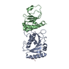
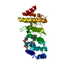
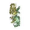
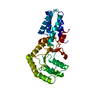
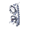
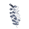
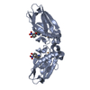
| Entry | Database: PDB / ID: 6d8v | ||||||
|---|---|---|---|---|---|---|---|
| Title | Methyl-accepting Chemotaxis protein X | ||||||
 Components Components | Probable chemoreceptor (Methyl-accepting chemotaxis) transmembrane protein | ||||||
 Keywords Keywords |  MEMBRANE PROTEIN / MCP / Chemotaxis MEMBRANE PROTEIN / MCP / Chemotaxis | ||||||
| Function / homology |  Function and homology informationchemotaxis / transmembrane signaling receptor activity / membrane => GO:0016020 / signal transduction Function and homology informationchemotaxis / transmembrane signaling receptor activity / membrane => GO:0016020 / signal transductionSimilarity search - Function | ||||||
| Biological species |  Rhizobium meliloti (bacteria) Rhizobium meliloti (bacteria) | ||||||
| Method | X-RAY DIFFRACTION / SYNCHROTRON / MOLECULAR REPLACEMENT / Resolution: 2.8 Å | ||||||
 Authors Authors | Shrestha, M. / Schubot, F.D. | ||||||
| Funding support |  United States, 1items United States, 1items
| ||||||
 Citation Citation | Journal: Biochem. J. / Year: 2018 Title: Structure of the sensory domain of McpX fromSinorhizobium meliloti, the first known bacterial chemotactic sensor for quaternary ammonium compounds. Authors: Shrestha, M. / Compton, K.K. / Mancl, J.M. / Webb, B.A. / Brown, A.M. / Scharf, B.E. / Schubot, F.D. | ||||||
| History |
|
- Structure visualization
| Structure viewer | Molecule: MolmilJmol/JSmol |
|---|
- Downloads & links
-Download
| PDBx/mmCIF format | 6d8v.cif.gz | 119.4 KB | Display | PDBx/mmCIF format |
|---|---|---|---|---|
| PDB format | pdb6d8v.ent.gz | 92.9 KB | Display | PDB format |
| PDBx/mmJSON format | 6d8v.json.gz | Tree view | PDBx/mmJSON format | |
| Others |  Other downloads Other downloads |
-Validation report
| Arichive directory | https://data.pdbj.org/pub/pdb/validation_reports/d8/6d8vftp://data.pdbj.org/pub/pdb/validation_reports/d8/6d8v | HTTPS FTP |
|---|
-Related structure data
| Similar structure data |
|---|

-Links
 PDBj
PDBj

- Assembly
| Deposited unit | 
| ||||||||
|---|---|---|---|---|---|---|---|---|---|
| 1 |
| ||||||||
| Unit cell |
|
-Components
| #1: Protein | Mass: 28718.098 Da / Num. of mol.: 1 Source method: isolated from a genetically manipulated source Source: (gene. exp.) Rhizobium meliloti (strain 1021) (bacteria)Strain: 1021 / Gene: mcpX, SMc01104 / Production host: Escherichia coli (E. coli) / References: UniProt: Q92SH9 |
|---|---|
| #2: Chemical | ChemComp-TRS / Tris  Mass: 122.143 Da / Num. of mol.: 1 / Source method: obtained synthetically / Formula: C4H12NO3 / Comment: pH buffer*YM Mass: 122.143 Da / Num. of mol.: 1 / Source method: obtained synthetically / Formula: C4H12NO3 / Comment: pH buffer*YM |
| #3: Chemical | ChemComp-PBE / 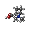  Mass: 144.192 Da / Num. of mol.: 1 / Source method: obtained synthetically / Formula: C7H14NO2 Mass: 144.192 Da / Num. of mol.: 1 / Source method: obtained synthetically / Formula: C7H14NO2 |
| #4: Water | ChemComp-HOH / Water Mass: 18.015 Da / Num. of mol.: 2 / Source method: isolated from a natural source / Formula: H2O Mass: 18.015 Da / Num. of mol.: 2 / Source method: isolated from a natural source / Formula: H2O |
-Experimental details
-Experiment
| Experiment | Method: X-RAY DIFFRACTION / Number of used crystals: 1 |
|---|
- Sample preparation
| Crystal | Density Matthews: 4.08 Å3/Da / Density % sol: 69.85 % |
|---|---|
| Crystal grow | Temperature: 296 K / Method: vapor diffusion, hanging drop Details: 0.08M Sodium acetate 1.6M Ammonium sulfate 20% glycerol |
-Data collection
| Diffraction | Mean temperature: 100 K |
|---|---|
| Diffraction source | Source: SYNCHROTRON / Site: APS / Beamline: 22-ID / Wavelength: 1 Å |
| Detector | Type: RAYONIX MX300-HS / Detector: CCD / Date: Jul 13, 2015 |
| Radiation | Protocol: SINGLE WAVELENGTH / Monochromatic (M) / Laue (L): M / Scattering type: x-ray |
| Radiation wavelength | Wavelength: 1 Å / Relative weight: 1 |
| Reflection | Resolution: 2.75→78.66 Å / Num. obs: 13433 / % possible obs: 99.7 % / Redundancy: 7 % / Net I/σ(I): 15.5 |
- Processing
| Software |
| ||||||||||||||||||||||||||||||||||||||||||
|---|---|---|---|---|---|---|---|---|---|---|---|---|---|---|---|---|---|---|---|---|---|---|---|---|---|---|---|---|---|---|---|---|---|---|---|---|---|---|---|---|---|---|---|
| Refinement | Method to determine structure: MOLECULAR REPLACEMENT / Resolution: 2.8→78.63 Å / SU ML: 0.41 / Cross valid method: THROUGHOUT / σ(F): 1.34 / Phase error: 28.02 / Stereochemistry target values: ML
| ||||||||||||||||||||||||||||||||||||||||||
| Solvent computation | Shrinkage radii: 0.9 Å / VDW probe radii: 1.11 Å / Solvent model: FLAT BULK SOLVENT MODEL | ||||||||||||||||||||||||||||||||||||||||||
| Displacement parameters | Biso max: 184.79 Å2 / Biso mean: 95.5076 Å2 / Biso min: 42.37 Å2 | ||||||||||||||||||||||||||||||||||||||||||
| Refinement step | Cycle: final / Resolution: 2.8→78.63 Å
| ||||||||||||||||||||||||||||||||||||||||||
| Refine LS restraints |
| ||||||||||||||||||||||||||||||||||||||||||
| LS refinement shell | Refine-ID: X-RAY DIFFRACTION / Rfactor Rfree error: 0 / Total num. of bins used: 5
| ||||||||||||||||||||||||||||||||||||||||||
| Refinement TLS params. | Method: refined / Origin x: 43.8551 Å / Origin y: 55.7719 Å / Origin z: 126.2002 Å
| ||||||||||||||||||||||||||||||||||||||||||
| Refinement TLS group |
|
