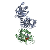
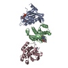
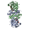
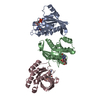
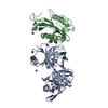
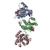
[English] 日本語
Yorodumi
- PDB-6d1v: Crystal structure of E. coli RppH-DapF complex, monomer bound to RNA -

+
Open data


ID or keywords:

Loading...

-
Basic information

Entry
Database: PDB / ID: 6d1v
TitleCrystal structure of E. coli RppH-DapF complex, monomer bound to RNA
Components
  • Diaminopimelate epimerase
  • RNA (5'-D(*(APC))-R(P*GP*U)-3')
  • RNA pyrophosphohydrolase
Keywordsisomerase/hydrolase/rna / RNA decay / RppH / DapF / isomerase-hydrolase complex / isomerase-hydrolase-rna complex
Function / homology
Function and homology information


diaminopimelate epimerase / diaminopimelate epimerase activity / RNA NAD-cap (NMN-forming) hydrolase activity / RNA decapping / mRNA 5'-diphosphatase activity / RNA destabilization / NAD-cap decapping / hydrolase activity, acting on acid anhydrides, in phosphorus-containing anhydrides / tRNA processing / lysine biosynthetic process via diaminopimelate ...diaminopimelate epimerase / diaminopimelate epimerase activity / RNA NAD-cap (NMN-forming) hydrolase activity / RNA decapping / mRNA 5'-diphosphatase activity / RNA destabilization / NAD-cap decapping / hydrolase activity, acting on acid anhydrides, in phosphorus-containing anhydrides / tRNA processing / lysine biosynthetic process via diaminopimelate / mRNA catabolic process / Hydrolases; Acting on acid anhydrides; In phosphorus-containing anhydrides / enzyme activator activity / magnesium ion binding / protein homodimerization activity / cytosol / cytoplasm
Similarity search - Function
RNA pyrophosphohydrolase RppH / Diaminopimelate epimerase, active site / Diaminopimelate epimerase signature. / Diaminopimelate epimerase, DapF / Diaminopimelate epimerase / Diaminopimelate Epimerase; Chain A, domain 1 / Diaminopimelate Epimerase; Chain A, domain 1 / NUDIX hydrolase / NUDIX hydrolase, conserved site / Nudix box signature. ...RNA pyrophosphohydrolase RppH / Diaminopimelate epimerase, active site / Diaminopimelate epimerase signature. / Diaminopimelate epimerase, DapF / Diaminopimelate epimerase / Diaminopimelate Epimerase; Chain A, domain 1 / Diaminopimelate Epimerase; Chain A, domain 1 / NUDIX hydrolase / NUDIX hydrolase, conserved site / Nudix box signature. / Nucleoside Triphosphate Pyrophosphohydrolase / Nucleoside Triphosphate Pyrophosphohydrolase / NUDIX domain / Nudix hydrolase domain profile. / NUDIX hydrolase domain / NUDIX hydrolase-like domain superfamily / Roll / Alpha-Beta Complex / Alpha Beta
Similarity search - Domain/homology
RNA / RNA pyrophosphohydrolase / Diaminopimelate epimerase / Diaminopimelate epimerase / RNA pyrophosphohydrolase
Similarity search - Component
Biological speciesEscherichia coli (E. coli)
MethodX-RAY DIFFRACTION / SYNCHROTRON / Resolution: 1.81 Å
AuthorsGao, A. / Serganov, A.
Funding support United States, 11items
OrganizationGrant numberCountry
National Institutes of Health/National Human Genome Research Institute (NIH/NHGRI)P41 GM103403 United States
National Institutes of Health/National Human Genome Research Institute (NIH/NHGRI)S10 RR029205 United States
Department of Energy (DOE, United States)DE-AC02-06CH11357 United States
Department of Energy (DOE, United States)KP1605010 United States
Department of Energy (DOE, United States)KC0401040 United States
National Institutes of Health/National Human Genome Research Institute (NIH/NHGRI)P41GM111244 United States
Department of Energy (DOE, United States)DE-SC0012704 United States
National Institutes of Health/National Human Genome Research Institute (NIH/NHGRI)R01GM112940 United States
National Institutes of Health/National Human Genome Research Institute (NIH/NHGRI)R01GM035769 United States
National Institutes of Health/National Human Genome Research Institute (NIH/NHGRI)R01AI108889 United States
National Institutes of Health/National Human Genome Research Institute (NIH/NHGRI)F99CA212474 United States
CitationJournal: Nucleic Acids Res. / Year: 2018
Title: Structural and kinetic insights into stimulation of RppH-dependent RNA degradation by the metabolic enzyme DapF.
Authors: Gao, A. / Vasilyev, N. / Luciano, D.J. / Levenson-Palmer, R. / Richards, J. / Marsiglia, W.M. / Traaseth, N.J. / Belasco, J.G. / Serganov, A.
History
DepositionApr 12, 2018Deposition site: RCSB / Processing site: RCSB
Revision 1.0May 23, 2018Provider: repository / Type: Initial release
Revision 1.1Aug 8, 2018Group: Data collection / Database references ...Data collection / Database references / Source and taxonomy / Structure summary
Category: citation / entity_name_com ...citation / entity_name_com / entity_src_gen / struct_ref / struct_ref_seq / struct_ref_seq_dif
Item: _citation.journal_volume / _citation.page_first ..._citation.journal_volume / _citation.page_first / _citation.page_last / _entity_name_com.name / _entity_src_gen.gene_src_strain / _entity_src_gen.pdbx_gene_src_gene / _entity_src_gen.pdbx_gene_src_ncbi_taxonomy_id / _entity_src_gen.pdbx_gene_src_scientific_name / _struct_ref.db_code / _struct_ref.pdbx_db_accession / _struct_ref.pdbx_seq_one_letter_code / _struct_ref_seq.db_align_end / _struct_ref_seq.pdbx_auth_seq_align_end / _struct_ref_seq.pdbx_db_accession / _struct_ref_seq.seq_align_end / _struct_ref_seq_dif.db_mon_id / _struct_ref_seq_dif.details / _struct_ref_seq_dif.pdbx_seq_db_accession_code / _struct_ref_seq_dif.pdbx_seq_db_seq_num
Revision 1.2Dec 4, 2019Group: Author supporting evidence / Category: pdbx_audit_support / Item: _pdbx_audit_support.funding_organization
Revision 1.3Mar 13, 2024Group: Data collection / Database references / Derived calculations
Category: chem_comp_atom / chem_comp_bond ...chem_comp_atom / chem_comp_bond / database_2 / pdbx_struct_conn_angle / struct_conn / struct_conn_type
Item: _database_2.pdbx_DOI / _database_2.pdbx_database_accession ..._database_2.pdbx_DOI / _database_2.pdbx_database_accession / _pdbx_struct_conn_angle.ptnr1_auth_asym_id / _pdbx_struct_conn_angle.ptnr1_auth_comp_id / _pdbx_struct_conn_angle.ptnr1_auth_seq_id / _pdbx_struct_conn_angle.ptnr1_label_asym_id / _pdbx_struct_conn_angle.ptnr1_label_atom_id / _pdbx_struct_conn_angle.ptnr1_label_comp_id / _pdbx_struct_conn_angle.ptnr1_label_seq_id / _pdbx_struct_conn_angle.ptnr2_auth_seq_id / _pdbx_struct_conn_angle.ptnr2_label_asym_id / _pdbx_struct_conn_angle.ptnr3_auth_asym_id / _pdbx_struct_conn_angle.ptnr3_auth_comp_id / _pdbx_struct_conn_angle.ptnr3_auth_seq_id / _pdbx_struct_conn_angle.ptnr3_label_asym_id / _pdbx_struct_conn_angle.ptnr3_label_atom_id / _pdbx_struct_conn_angle.ptnr3_label_comp_id / _pdbx_struct_conn_angle.ptnr3_label_seq_id / _pdbx_struct_conn_angle.value / _struct_conn.conn_type_id / _struct_conn.id / _struct_conn.pdbx_dist_value / _struct_conn.pdbx_leaving_atom_flag / _struct_conn.ptnr1_auth_asym_id / _struct_conn.ptnr1_auth_comp_id / _struct_conn.ptnr1_auth_seq_id / _struct_conn.ptnr1_label_asym_id / _struct_conn.ptnr1_label_atom_id / _struct_conn.ptnr1_label_comp_id / _struct_conn.ptnr1_label_seq_id / _struct_conn.ptnr2_auth_asym_id / _struct_conn.ptnr2_auth_comp_id / _struct_conn.ptnr2_auth_seq_id / _struct_conn.ptnr2_label_asym_id / _struct_conn.ptnr2_label_atom_id / _struct_conn.ptnr2_label_comp_id / _struct_conn.ptnr2_label_seq_id / _struct_conn_type.id

-
Structure visualization

Structure viewerMolecule:
MolmilJmol/JSmol

Downloads & links

-
Assembly

Deposited unit
A: Diaminopimelate epimerase
B: RNA pyrophosphohydrolase
C: RNA (5'-D(*(APC))-R(P*GP*U)-3')
hetero molecules


Theoretical massNumber of molelcules
Total (without water)51,21618
Polymers50,3213
Non-polymers89515
Water5,188288
1


  • Idetical with deposited unit
  • defined by author
  • Evidence: gel filtration
TypeNameSymmetry operationNumber
identity operation1_555x,y,z1
Unit cell
Length a, b, c (Å)160.644, 191.986, 51.207
Angle α, β, γ (deg.)90.00, 90.00, 90.00
Int Tables number20
Space group name H-MC2221
Components on special symmetry positions
IDModelComponents
11B-207-

CL

-
Components

-
Protein , 2 types, 2 molecules AB

#1: Protein Diaminopimelate epimerase /


Mass: 30332.629 Da / Num. of mol.: 1
Source method: isolated from a genetically manipulated source
Source: (gene. exp.) Escherichia coli (E. coli) / Strain: K12 / Gene: dapF / Production host: Escherichia coli (E. coli) / References: UniProt: P0A6K1, UniProt: A7ZU14*PLUS
#2: Protein RNA pyrophosphohydrolase / (Di)nucleoside polyphosphate hydrolase / Ap5A pyrophosphatase


Mass: 18894.695 Da / Num. of mol.: 1
Source method: isolated from a genetically manipulated source
Source: (gene. exp.) Escherichia coli (E. coli) / Strain: K12 / Gene: rppH, nudH, ygdP, b2830, JW2798 / Production host: Escherichia coli (E. coli)
References: UniProt: P0A776, UniProt: A7ZQT6*PLUS, Hydrolases; Acting on acid anhydrides; In phosphorus-containing anhydrides

-
RNA chain , 1 types, 1 molecules C

#3: RNA chain RNA (5'-D(*(APC))-R(P*GP*U)-3')


Mass: 1093.607 Da / Num. of mol.: 1 / Source method: obtained synthetically / Source: (synth.) Escherichia coli (E. coli)

-
Non-polymers , 4 types, 303 molecules

#4: Chemical
ChemComp-GOL / GLYCEROL / GLYCERIN / PROPANE-1,2,3-TRIOL / Glycerol


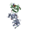
Mass: 92.094 Da / Num. of mol.: 7 / Source method: obtained synthetically / Formula: C3H8O3
#5: Chemical
ChemComp-CL / CHLORIDE ION / Chloride


Mass: 35.453 Da / Num. of mol.: 5 / Source method: obtained synthetically / Formula: Cl
#6: Chemical ChemComp-MG / MAGNESIUM ION


Mass: 24.305 Da / Num. of mol.: 3 / Source method: obtained synthetically / Formula: Mg
#7: Water ChemComp-HOH / water / Water


Mass: 18.015 Da / Num. of mol.: 288 / Source method: isolated from a natural source / Formula: H2O

-
Experimental details

-
Experiment

ExperimentMethod: X-RAY DIFFRACTION / Number of used crystals: 1

-
Sample preparation

CrystalDensity Matthews: 3.92 Å3/Da / Density % sol: 68.65 %
Crystal growTemperature: 292 K / Method: vapor diffusion / Details: 30% (v/v) PEG400 and 0.1 M CHES, pH 9.2

-
Data collection

DiffractionMean temperature: 100 K
Diffraction sourceSource: SYNCHROTRON / Site: APS / Beamline: 24-ID-C / Wavelength: 0.9791 Å
DetectorType: ADSC QUANTUM 315 / Detector: CCD / Date: Mar 19, 2016
RadiationProtocol: SINGLE WAVELENGTH / Monochromatic (M) / Laue (L): M / Scattering type: x-ray
Radiation wavelengthWavelength: 0.9791 Å / Relative weight: 1
ReflectionResolution: 1.81→123.2 Å / Num. obs: 70344 / % possible obs: 96.8 % / Redundancy: 2.9 % / Rmerge(I) obs: 0.037 / Net I/σ(I): 12.6
Reflection shellResolution: 1.81→1.85 Å / Rmerge(I) obs: 0.989 / Num. unique all: 3453

-
Processing

Software
NameVersionClassification
PHENIX(1.11.1_2575: ???)refinement
Cootmodel building
XDSdata scaling
PHENIXphasing
RefinementResolution: 1.81→61.601 Å / SU ML: 0.26 / Cross valid method: FREE R-VALUE / σ(F): 1.35 / Phase error: 24.1
RfactorNum. reflection% reflection
Rfree0.2196 2000 2.84 %
Rwork0.1988 --
obs0.1994 70302 96.78 %
Solvent computationShrinkage radii: 0.9 Å / VDW probe radii: 1.11 Å
Refinement stepCycle: LAST / Resolution: 1.81→61.601 Å
ProteinNucleic acidLigandSolventTotal
Num. atoms3438 58 49 289 3834
Refine LS restraints
Refine-IDTypeDev idealNumber
X-RAY DIFFRACTIONf_bond_d0.0083630
X-RAY DIFFRACTIONf_angle_d1.0364918
X-RAY DIFFRACTIONf_dihedral_angle_d5.8382970
X-RAY DIFFRACTIONf_chiral_restr0.058520
X-RAY DIFFRACTIONf_plane_restr0.006633
LS refinement shell
Resolution (Å)Rfactor RfreeNum. reflection RfreeRfactor RworkNum. reflection RworkRefine-ID% reflection obs (%)
1.8104-1.85560.36931210.33994139X-RAY DIFFRACTION83
1.8556-1.90580.36241450.3154977X-RAY DIFFRACTION99
1.9058-1.96190.33151460.28154931X-RAY DIFFRACTION99
1.9619-2.02520.27671440.25654936X-RAY DIFFRACTION99
2.0252-2.09760.25541440.24964942X-RAY DIFFRACTION99
2.0976-2.18160.26741450.22974945X-RAY DIFFRACTION99
2.1816-2.28090.24981450.22284963X-RAY DIFFRACTION99
2.2809-2.40110.29341450.22694926X-RAY DIFFRACTION99
2.4011-2.55160.26891460.21674989X-RAY DIFFRACTION99
2.5516-2.74860.26491430.22154877X-RAY DIFFRACTION97
2.7486-3.02520.21641440.20974915X-RAY DIFFRACTION98
3.0252-3.46290.23141420.19954872X-RAY DIFFRACTION96
3.4629-4.36270.18091420.16764865X-RAY DIFFRACTION95
4.3627-61.63710.1711480.16615025X-RAY DIFFRACTION94

+
About Yorodumi

-
News

-
Feb 9, 2022. New format data for meta-information of EMDB entries

New format data for meta-information of EMDB entries

  • Version 3 of the EMDB header file is now the official format.
  • The previous official version 1.9 will be removed from the archive.

Related info.:EMDB header

External links:wwPDB to switch to version 3 of the EMDB data model

-
Aug 12, 2020. Covid-19 info

Covid-19 info

URL: https://pdbj.org/emnavi/covid19.php

New page: Covid-19 featured information page in EM Navigator.

Related info.:Covid-19 info / Mar 5, 2020. Novel coronavirus structure data

+
Mar 5, 2020. Novel coronavirus structure data

Novel coronavirus structure data

Related info.:Yorodumi Speices / Aug 12, 2020. Covid-19 info

External links:COVID-19 featured content - PDBj / Molecule of the Month (242):Coronavirus Proteases

+
Jan 31, 2019. EMDB accession codes are about to change! (news from PDBe EMDB page)

EMDB accession codes are about to change! (news from PDBe EMDB page)

  • The allocation of 4 digits for EMDB accession codes will soon come to an end. Whilst these codes will remain in use, new EMDB accession codes will include an additional digit and will expand incrementally as the available range of codes is exhausted. The current 4-digit format prefixed with “EMD-” (i.e. EMD-XXXX) will advance to a 5-digit format (i.e. EMD-XXXXX), and so on. It is currently estimated that the 4-digit codes will be depleted around Spring 2019, at which point the 5-digit format will come into force.
  • The EM Navigator/Yorodumi systems omit the EMD- prefix.

Related info.:Q: What is EMD? / ID/Accession-code notation in Yorodumi/EM Navigator

External links:EMDB Accession Codes are Changing Soon! / Contact to PDBj

+
Jul 12, 2017. Major update of PDB

Major update of PDB

  • wwPDB released updated PDB data conforming to the new PDBx/mmCIF dictionary.
  • This is a major update changing the version number from 4 to 5, and with Remediation, in which all the entries are updated.
  • In this update, many items about electron microscopy experimental information are reorganized (e.g. em_software).
  • Now, EM Navigator and Yorodumi are based on the updated data.

External links:wwPDB Remediation / Enriched Model Files Conforming to OneDep Data Standards Now Available in the PDB FTP Archive

-
Yorodumi

Thousand views of thousand structures

  • Yorodumi is a browser for structure data from EMDB, PDB, SASBDB, etc.
  • This page is also the successor to EM Navigator detail page, and also detail information page/front-end page for Omokage search.
  • The word "yorodu" (or yorozu) is an old Japanese word meaning "ten thousand". "mi" (miru) is to see.

Related info.:EMDB / PDB / SASBDB / Comparison of 3 databanks / Yorodumi Search / Aug 31, 2016. New EM Navigator & Yorodumi / Yorodumi Papers / Jmol/JSmol / Function and homology information / Changes in new EM Navigator and Yorodumi

Read more