 Movie
Movie Controller

[English] 日本語
 Yorodumi
- PDB-6d0n: Crystal structure of a CLC-type fluoride/proton antiporter, V319G... -
+ Open data

- Basic information
| Entry | Database: PDB / ID: 6d0n | |||||||||
|---|---|---|---|---|---|---|---|---|---|---|
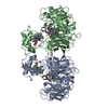
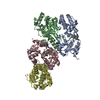
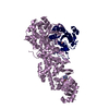
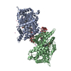
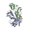
| Title | Crystal structure of a CLC-type fluoride/proton antiporter, V319G mutant | |||||||||
 Components Components |
| |||||||||
 Keywords Keywords |  TRANSPORT PROTEIN / fluoride/proton antiporter / CLC membrane protein TRANSPORT PROTEIN / fluoride/proton antiporter / CLC membrane protein | |||||||||
| Function / homology | Chloride channel, voltage gated / Chloride channel, core / Voltage gated chloride channel / voltage-gated chloride channel activity / membrane / FLUORIDE ION / (CARBAMOYLMETHYL-CARBOXYMETHYL-AMINO)-ACETIC ACID / Voltage-gated chloride channel protein Function and homology information Function and homology information | |||||||||
| Biological species |  Enterococcus casseliflavus (bacteria) Enterococcus casseliflavus (bacteria)synthetic construct (others) | |||||||||
| Method | X-RAY DIFFRACTION / SYNCHROTRON / MOLECULAR REPLACEMENT / Resolution: 3.12 Å | |||||||||
 Authors Authors | Last, N.B. / Stockbridge, R.B. / Wilson, A.E. / Shane, T. / Kolmakova-Partensky, L. / Koide, A. / Koide, S. / Miller, C. | |||||||||
| Funding support |  United States, 2items United States, 2items
| |||||||||
 Citation Citation | Journal: Nat. Struct. Mol. Biol. / Year: 2018 Title: A CLC-type F-/H+antiporter in ion-swapped conformations. Authors: Last, N.B. / Stockbridge, R.B. / Wilson, A.E. / Shane, T. / Kolmakova-Partensky, L. / Koide, A. / Koide, S. / Miller, C. | |||||||||
| History |
|
- Structure visualization
| Structure viewer | Molecule: MolmilJmol/JSmol |
|---|
- Downloads & links
-Download
| PDBx/mmCIF format | 6d0n.cif.gz | 471.3 KB | Display | PDBx/mmCIF format |
|---|---|---|---|---|
| PDB format | pdb6d0n.ent.gz | 332 KB | Display | PDB format |
| PDBx/mmJSON format | 6d0n.json.gz | Tree view | PDBx/mmJSON format | |
| Others |  Other downloads Other downloads |
-Validation report
| Arichive directory | https://data.pdbj.org/pub/pdb/validation_reports/d0/6d0nftp://data.pdbj.org/pub/pdb/validation_reports/d0/6d0n | HTTPS FTP |
|---|
-Related structure data
| Related structure data |  6d0jSC 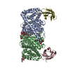 6d0kC S: Starting model for refinement C: citing same article ( |
|---|---|
| Similar structure data |


-Links
 PDBj
PDBj

- Assembly
| Deposited unit | 
| |||||||||||||||||||||||||||||||||||||||||||||||||||||||
|---|---|---|---|---|---|---|---|---|---|---|---|---|---|---|---|---|---|---|---|---|---|---|---|---|---|---|---|---|---|---|---|---|---|---|---|---|---|---|---|---|---|---|---|---|---|---|---|---|---|---|---|---|---|---|---|---|
| 1 |
| |||||||||||||||||||||||||||||||||||||||||||||||||||||||
| Unit cell |
| |||||||||||||||||||||||||||||||||||||||||||||||||||||||
| Noncrystallographic symmetry (NCS) | NCS domain:
NCS domain segments:
NCS ensembles :
|
-Components
-Protein / Antibody / Sugars , 3 types, 10 molecules ABDC
| #1: Protein | Mass: 45626.676 Da / Num. of mol.: 2 / Mutation: M4I, V319G Source method: isolated from a genetically manipulated source Source: (gene. exp.) Enterococcus casseliflavus (strain EC10) (bacteria)Strain: EC10 / Gene: ECAG_02710 / Production host: Escherichia coli (E. coli) / Strain (production host): BL21(DE3) / References: UniProt: C9CPP6#2: Antibody | Mass: 9891.871 Da / Num. of mol.: 2 Source method: isolated from a genetically manipulated source Source: (gene. exp.) synthetic construct (others) / Production host: Escherichia coli (E. coli) / Strain (production host): BL21(DE3)#3: Sugar | ChemComp-DMU /  Type: D-saccharide / Mass: 482.562 Da / Num. of mol.: 6 Type: D-saccharide / Mass: 482.562 Da / Num. of mol.: 6Source method: isolated from a genetically manipulated source Formula: C22H42O11 / Comment: detergent*YM |
|---|
-Non-polymers , 3 types, 13 molecules 


| #4: Chemical | ChemComp-F / Fluoride Mass: 18.998 Da / Num. of mol.: 1 / Source method: obtained synthetically / Formula: F Mass: 18.998 Da / Num. of mol.: 1 / Source method: obtained synthetically / Formula: F |
|---|---|
| #5: Chemical | ChemComp-MHA / (ADA (buffer) Mass: 190.154 Da / Num. of mol.: 1 / Source method: obtained synthetically / Formula: C6H10N2O5 / Comment: pH buffer*YM Mass: 190.154 Da / Num. of mol.: 1 / Source method: obtained synthetically / Formula: C6H10N2O5 / Comment: pH buffer*YM |
| #6: Water | ChemComp-HOH / WaterMass: 18.015 Da / Num. of mol.: 11 / Source method: isolated from a natural source / Formula: H2O |
-Experimental details
-Experiment
| Experiment | Method: X-RAY DIFFRACTION / Number of used crystals: 1 |
|---|
- Sample preparation
| Crystal | Density Matthews: 4.77 Å3/Da / Density % sol: 74.2 % |
|---|---|
| Crystal grow | Temperature: 295 K / Method: vapor diffusion, sitting drop / pH: 6.2 Details: 10 mM HEPES, pH 7.0 100 mM ADA, pH 6.2 100 mM NaF 100 mM K Formate 24% PEG 600 |
-Data collection
| Diffraction | Mean temperature: 100 K |
|---|---|
| Diffraction source | Source: SYNCHROTRON / Site: ALS / Beamline: 5.0.2 / Wavelength: 1 Å |
| Detector | Type: DECTRIS PILATUS3 6M / Detector: PIXEL / Date: Oct 29, 2017 |
| Radiation | Monochromator: Double-crystal, Si(111) / Protocol: SINGLE WAVELENGTH / Monochromatic (M) / Laue (L): M / Scattering type: x-ray |
| Radiation wavelength | Wavelength: 1 Å / Relative weight: 1 |
| Reflection | Resolution: 3.12→49.67 Å / Num. obs: 36328 / % possible obs: 99.9 % / Redundancy: 17.6 % / Biso Wilson estimate: 124.36 Å2 / CC1/2: 1 / Rmerge(I) obs: 0.082 / Net I/σ(I): 12.2 |
| Reflection shell | Resolution: 3.12→3.26 Å / Redundancy: 17.3 % / Rmerge(I) obs: 2.81 / Mean I/σ(I) obs: 1.3 / Num. unique obs: 4328 / CC1/2: 0.699 / % possible all: 99.6 |
- Processing
| Software |
| ||||||||||||||||||||||||||||||||||||||||||||||||||||||||||||||||||||||||||||||||||||||||||||||||||
|---|---|---|---|---|---|---|---|---|---|---|---|---|---|---|---|---|---|---|---|---|---|---|---|---|---|---|---|---|---|---|---|---|---|---|---|---|---|---|---|---|---|---|---|---|---|---|---|---|---|---|---|---|---|---|---|---|---|---|---|---|---|---|---|---|---|---|---|---|---|---|---|---|---|---|---|---|---|---|---|---|---|---|---|---|---|---|---|---|---|---|---|---|---|---|---|---|---|---|---|
| Refinement | Method to determine structure: MOLECULAR REPLACEMENT Starting model: 6D0J Resolution: 3.12→49.67 Å / SU ML: 0.5165 / Cross valid method: FREE R-VALUE / σ(F): 1.35 / Phase error: 37.6874 / Stereochemistry target values: GeoStd + Monomer Library
| ||||||||||||||||||||||||||||||||||||||||||||||||||||||||||||||||||||||||||||||||||||||||||||||||||
| Solvent computation | Shrinkage radii: 0.9 Å / VDW probe radii: 1.11 Å / Solvent model: FLAT BULK SOLVENT MODEL | ||||||||||||||||||||||||||||||||||||||||||||||||||||||||||||||||||||||||||||||||||||||||||||||||||
| Displacement parameters | Biso mean: 169.94 Å2 | ||||||||||||||||||||||||||||||||||||||||||||||||||||||||||||||||||||||||||||||||||||||||||||||||||
| Refinement step | Cycle: LAST / Resolution: 3.12→49.67 Å
| ||||||||||||||||||||||||||||||||||||||||||||||||||||||||||||||||||||||||||||||||||||||||||||||||||
| Refine LS restraints |
| ||||||||||||||||||||||||||||||||||||||||||||||||||||||||||||||||||||||||||||||||||||||||||||||||||
| LS refinement shell |
| ||||||||||||||||||||||||||||||||||||||||||||||||||||||||||||||||||||||||||||||||||||||||||||||||||
| Refinement TLS params. | Method: refined / Origin x: 137.770267351 Å / Origin y: 159.593966428 Å / Origin z: 1.70189385981 Å
| ||||||||||||||||||||||||||||||||||||||||||||||||||||||||||||||||||||||||||||||||||||||||||||||||||
| Refinement TLS group | Selection details: all |
