 Movie
Movie Controller

[English] 日本語
 Yorodumi
- PDB-6cu5: Crystal structure of a protein arginine N-methyltransferase from ... -
+ Open data

- Basic information
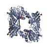
| Entry | Database: PDB / ID: 6cu5 | ||||||
|---|---|---|---|---|---|---|---|
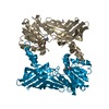
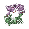
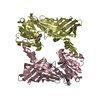
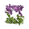
| Title | Crystal structure of a protein arginine N-methyltransferase from Naegleria fowleri bound to SAH | ||||||
 Components Components | protein arginine N-methyltransferase | ||||||
 Keywords Keywords |  TRANSFERASE / NIAID / structural genomics / brain eating amoeba / Primary Amebic Meningoencephalitis (PAM) / Amebic Encephalitis / Seattle Structural Genomics Center for Infectious Disease / SSGCID TRANSFERASE / NIAID / structural genomics / brain eating amoeba / Primary Amebic Meningoencephalitis (PAM) / Amebic Encephalitis / Seattle Structural Genomics Center for Infectious Disease / SSGCID | ||||||
| Function / homology |  Function and homology information Function and homology informationpeptidyl-arginine methylation / protein-arginine N-methyltransferase activity Similarity search - Function | ||||||
| Biological species |  Naegleria fowleri (brain-eating amoeba) Naegleria fowleri (brain-eating amoeba) | ||||||
| Method | X-RAY DIFFRACTION / SYNCHROTRON / MOLECULAR REPLACEMENT / molecular replacement / Resolution: 2.7 Å | ||||||
 Authors Authors | Seattle Structural Genomics Center for Infectious Disease (SSGCID) | ||||||
 Citation Citation | Journal: To Be Published Title: Crystal structure of a protein arginine N-methyltransferase from Naegleria fowleri bound to SAH Authors: Edwards, T.E. / Dranow, D.M. / Lorimer, D.D. / Horanyi, P.S. | ||||||
| History |
|
- Structure visualization
| Structure viewer | Molecule: MolmilJmol/JSmol |
|---|
- Downloads & links
-Download
| PDBx/mmCIF format | 6cu5.cif.gz | 261.4 KB | Display | PDBx/mmCIF format |
|---|---|---|---|---|
| PDB format | pdb6cu5.ent.gz | 209.6 KB | Display | PDB format |
| PDBx/mmJSON format | 6cu5.json.gz | Tree view | PDBx/mmJSON format | |
| Others |  Other downloads Other downloads |
-Validation report
| Arichive directory | https://data.pdbj.org/pub/pdb/validation_reports/cu/6cu5ftp://data.pdbj.org/pub/pdb/validation_reports/cu/6cu5 | HTTPS FTP |
|---|
-Related structure data
| Related structure data | 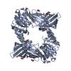 6cu3S S: Starting model for refinement |
|---|---|
| Similar structure data | |
| Other databases |


-Links
 PDBj
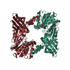
PDBj- Assembly
| Deposited unit | 
| ||||||||||||||||||||||||||||||||||||||||||||||||||||||||||||||||||||||||||||||||||||||||||
|---|---|---|---|---|---|---|---|---|---|---|---|---|---|---|---|---|---|---|---|---|---|---|---|---|---|---|---|---|---|---|---|---|---|---|---|---|---|---|---|---|---|---|---|---|---|---|---|---|---|---|---|---|---|---|---|---|---|---|---|---|---|---|---|---|---|---|---|---|---|---|---|---|---|---|---|---|---|---|---|---|---|---|---|---|---|---|---|---|---|---|---|
| 1 |
| ||||||||||||||||||||||||||||||||||||||||||||||||||||||||||||||||||||||||||||||||||||||||||
| Unit cell |
| ||||||||||||||||||||||||||||||||||||||||||||||||||||||||||||||||||||||||||||||||||||||||||
| Noncrystallographic symmetry (NCS) | NCS domain:
NCS domain segments: Ens-ID: 1
|