 Movie
Movie Controller

[English] 日本語
 Yorodumi
- PDB-6cqi: 2.42A Crystal structure of Mycobacterium tuberculosis Topoisomera... -
+ Open data

- Basic information
| Entry | Database: PDB / ID: 6cqi | ||||||||||||
|---|---|---|---|---|---|---|---|---|---|---|---|---|---|
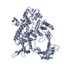
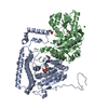
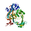
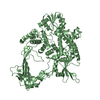
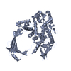
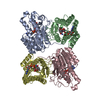
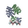
| Title | 2.42A Crystal structure of Mycobacterium tuberculosis Topoisomerase I in complex with an oligonucleotide MTS2-11 | ||||||||||||
 Components Components |
| ||||||||||||
 Keywords Keywords | ISOMERASE/DNA /  Mycobacterium tuberculosis / Topoisomerase I / co-crystal / complex with oligonucleotide / Isomerase-DNA complex / ISOMERASE Mycobacterium tuberculosis / Topoisomerase I / co-crystal / complex with oligonucleotide / Isomerase-DNA complex / ISOMERASE | ||||||||||||
| Function / homology |  Function and homology information Function and homology informationnegative regulation of ribonuclease activity / DNA topoisomerase / DNA topoisomerase type I (single strand cut, ATP-independent) activity / DNA topological change / peptidoglycan-based cell wall / magnesium ion binding / DNA binding / plasma membrane / cytosolSimilarity search - Function | ||||||||||||
| Biological species |   Mycobacterium tuberculosis (bacteria) Mycobacterium tuberculosis (bacteria) | ||||||||||||
| Method | X-RAY DIFFRACTION / SYNCHROTRON / MOLECULAR REPLACEMENT / Resolution: 2.42 Å | ||||||||||||
 Authors Authors | Cao, N. / Thirunavukkarasu, A. / Tan, K. / Tse-Dinh, Y.-C. | ||||||||||||
| Funding support |  United States, 3items United States, 3items
| ||||||||||||
 Citation Citation | Journal: Nucleic Acids Res. / Year: 2018 Title: Investigating mycobacterial topoisomerase I mechanism from the analysis of metal and DNA substrate interactions at the active site. Authors: Cao, N. / Tan, K. / Annamalai, T. / Joachimiak, A. / Tse-Dinh, Y.C. | ||||||||||||
| History |
|
- Structure visualization
| Structure viewer | Molecule: MolmilJmol/JSmol |
|---|
- Downloads & links
-Download
| PDBx/mmCIF format | 6cqi.cif.gz | 290.3 KB | Display | PDBx/mmCIF format |
|---|---|---|---|---|
| PDB format | pdb6cqi.ent.gz | 230 KB | Display | PDB format |
| PDBx/mmJSON format | 6cqi.json.gz | Tree view | PDBx/mmJSON format | |
| Others |  Other downloads Other downloads |
-Validation report
| Arichive directory | https://data.pdbj.org/pub/pdb/validation_reports/cq/6cqiftp://data.pdbj.org/pub/pdb/validation_reports/cq/6cqi | HTTPS FTP |
|---|
-Related structure data
| Related structure data | 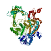 5uj1C  5ujyC  6cq2C  5ukt S: Starting model for refinement C: citing same article ( |
|---|---|
| Similar structure data |


-Links
 PDBj
PDBj


- Assembly
| Deposited unit | 
| ||||||||
|---|---|---|---|---|---|---|---|---|---|
| 1 |
| ||||||||
| Unit cell |
|
-Components
-Protein / DNA chain , 2 types, 2 molecules AB
| #1: Protein | Topoisomerase / DNA topoisomerase I / Omega-protein / Relaxing enzyme / Swivelase / Untwisting enzyme Mass: 77843.500 Da / Num. of mol.: 1 Source method: isolated from a genetically manipulated source Source: (gene. exp.) Mycobacterium tuberculosis (strain ATCC 25618 / H37Rv) (bacteria)Strain: ATCC 25618 / H37Rv / Gene: topA, Rv3646c, MTCY15C10.06 / Plasmid: PET-HIS6-MOCR TEV-LIC / Production host: Escherichia coli (E. coli) / Strain (production host): T7 EXPRESS CRYSTAL / References: UniProt: P9WG49, DNA topoisomerase |
|---|---|
| #2: DNA chain | Mass: 3300.159 Da / Num. of mol.: 1 / Source method: obtained synthetically / Source: (synth.) Mycobacterium tuberculosis (bacteria) |
-Non-polymers , 4 types, 24 molecules 


| #3: Chemical | Formic acid Mass: 46.025 Da / Num. of mol.: 2 / Source method: obtained synthetically / Formula: CH2O2 Mass: 46.025 Da / Num. of mol.: 2 / Source method: obtained synthetically / Formula: CH2O2#4: Chemical | ChemComp-GOL / | Glycerol Mass: 92.094 Da / Num. of mol.: 1 / Source method: obtained synthetically / Formula: C3H8O3 Mass: 92.094 Da / Num. of mol.: 1 / Source method: obtained synthetically / Formula: C3H8O3#5: Chemical | Acetate Mass: 59.044 Da / Num. of mol.: 3 / Source method: obtained synthetically / Formula: C2H3O2 Mass: 59.044 Da / Num. of mol.: 3 / Source method: obtained synthetically / Formula: C2H3O2#6: Water | ChemComp-HOH / | WaterMass: 18.015 Da / Num. of mol.: 18 / Source method: isolated from a natural source / Formula: H2O |
|---|
-Experimental details
-Experiment
| Experiment | Method: X-RAY DIFFRACTION / Number of used crystals: 1 |
|---|
- Sample preparation
| Crystal | Density Matthews: 2.46 Å3/Da / Density % sol: 50.06 % |
|---|---|
| Crystal grow | Temperature: 289 K / Method: vapor diffusion, sitting drop / pH: 8 Details: 0.2 M ammonium phosphate dibasic, 20% w/v Polyethylene glycol 3,350 |
-Data collection
| Diffraction | Mean temperature: 100 K |
|---|---|
| Diffraction source | Source: SYNCHROTRON / Site: APS / Beamline: 19-ID / Wavelength: 0.97918 Å |
| Detector | Type: DECTRIS PILATUS3 S 6M / Detector: PIXEL / Date: Mar 4, 2017 / Details: mirror |
| Radiation | Monochromator: silicon 111 / Protocol: SINGLE WAVELENGTH / Monochromatic (M) / Laue (L): M / Scattering type: x-ray |
| Radiation wavelength | Wavelength: 0.97918 Å / Relative weight: 1 |
| Reflection | Resolution: 2.42→47 Å / Num. obs: 29799 / % possible obs: 97.1 % / Observed criterion σ(I): -3 / Redundancy: 4 % / Biso Wilson estimate: 55.9 Å2 / Rmerge(I) obs: 0.087 / Rpim(I) all: 0.047 / Χ2: 1.25 / Net I/σ(I): 19.5 |
| Reflection shell | Resolution: 2.42→2.46 Å / Redundancy: 3.2 % / Rmerge(I) obs: 0.641 / Mean I/σ(I) obs: 1.2 / Num. unique obs: 1268 / CC1/2: 0.818 / Rpim(I) all: 0.368 / Χ2: 0.642 / % possible all: 82.4 |
- Processing
| Software |
| |||||||||||||||||||||||||||||||||||||||||||||||||||||||||||||||||||||||||||||||||||||||||||||||||||||||||||||||||||||||||||||
|---|---|---|---|---|---|---|---|---|---|---|---|---|---|---|---|---|---|---|---|---|---|---|---|---|---|---|---|---|---|---|---|---|---|---|---|---|---|---|---|---|---|---|---|---|---|---|---|---|---|---|---|---|---|---|---|---|---|---|---|---|---|---|---|---|---|---|---|---|---|---|---|---|---|---|---|---|---|---|---|---|---|---|---|---|---|---|---|---|---|---|---|---|---|---|---|---|---|---|---|---|---|---|---|---|---|---|---|---|---|---|---|---|---|---|---|---|---|---|---|---|---|---|---|---|---|---|
| Refinement | Method to determine structure: MOLECULAR REPLACEMENT Starting model: 5UKT 5ukt Resolution: 2.42→47 Å / SU ML: 0.37 / Cross valid method: FREE R-VALUE / σ(F): 1.34 / Phase error: 34.41 / Stereochemistry target values: ML
| |||||||||||||||||||||||||||||||||||||||||||||||||||||||||||||||||||||||||||||||||||||||||||||||||||||||||||||||||||||||||||||
| Solvent computation | Shrinkage radii: 0.9 Å / VDW probe radii: 1.11 Å / Solvent model: FLAT BULK SOLVENT MODEL | |||||||||||||||||||||||||||||||||||||||||||||||||||||||||||||||||||||||||||||||||||||||||||||||||||||||||||||||||||||||||||||
| Refinement step | Cycle: LAST / Resolution: 2.42→47 Å
| |||||||||||||||||||||||||||||||||||||||||||||||||||||||||||||||||||||||||||||||||||||||||||||||||||||||||||||||||||||||||||||
| Refine LS restraints |
| |||||||||||||||||||||||||||||||||||||||||||||||||||||||||||||||||||||||||||||||||||||||||||||||||||||||||||||||||||||||||||||
| LS refinement shell |
| |||||||||||||||||||||||||||||||||||||||||||||||||||||||||||||||||||||||||||||||||||||||||||||||||||||||||||||||||||||||||||||
| Refinement TLS params. | Method: refined / Refine-ID: X-RAY DIFFRACTION
| |||||||||||||||||||||||||||||||||||||||||||||||||||||||||||||||||||||||||||||||||||||||||||||||||||||||||||||||||||||||||||||
| Refinement TLS group |
|
