 Movie
Movie Controller

[English] 日本語
 Yorodumi
- PDB-6cfp: CRYSTAL STRUCTURE OF POLYMERASE ACID PROTEIN (PA) FROM INFLUENZA ... -
+ Open data

- Basic information
| Entry | Database: PDB / ID: 6cfp | ||||||
|---|---|---|---|---|---|---|---|
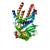
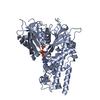
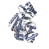
| Title | CRYSTAL STRUCTURE OF POLYMERASE ACID PROTEIN (PA) FROM INFLUENZA A VIRUS, WILSON-SMITH/1933 (H1N1) BOUND TO FRAGMENT HIT BSI-70565 1-{1-[4-FLUOROPHENYL)METHYL]-2-METHYL-1H-IMIDAZOL-4-YL}ETHAN-1-ONE | ||||||
 Components Components | Ubiquitin-like protein SMT3,Polymerase acidic protein | ||||||
 Keywords Keywords |  VIRAL PROTEIN / INFLUENZA / H1N1 / FRAGMENT-BASED DRUG DISCOVERY / Structural Genomics / Seattle Structural Genomics Center for Infectious Disease / SSGCID VIRAL PROTEIN / INFLUENZA / H1N1 / FRAGMENT-BASED DRUG DISCOVERY / Structural Genomics / Seattle Structural Genomics Center for Infectious Disease / SSGCID | ||||||
| Function / homology |  Function and homology informationRNA polymerase activity / SUMO is conjugated to E1 (UBA2:SAE1) / SUMOylation of nuclear envelope proteins / SUMO is transferred from E1 to E2 (UBE2I, UBC9) / SUMOylation of SUMOylation proteins / SUMO is proteolytically processed / SUMOylation of transcription factors / Postmitotic nuclear pore complex (NPC) reformation / SUMOylation of transcription cofactors / septin ring ...RNA polymerase activity / SUMO is conjugated to E1 (UBA2:SAE1) / SUMOylation of nuclear envelope proteins / SUMO is transferred from E1 to E2 (UBE2I, UBC9) / SUMOylation of SUMOylation proteins / SUMO is proteolytically processed / SUMOylation of transcription factors / Postmitotic nuclear pore complex (NPC) reformation / SUMOylation of transcription cofactors / septin ring / SUMOylation of DNA damage response and repair proteins / SUMOylation of RNA binding proteins / SUMOylation of DNA replication proteins / negative regulation of viral transcription / SUMOylation of chromatin organization proteins / negative stranded viral RNA replication / negative regulation of viral genome replication / cap snatching / ubiquitin-like protein ligase binding / symbiont-mediated suppression of host mRNA transcription via inhibition of RNA polymerase II activity / protein sumoylation / condensed nuclear chromosome / protein tag activity / protein import into nucleus / endonuclease activity / host cell cytoplasm / Hydrolases; Acting on ester bonds / DNA-templated transcription / host cell nucleus / RNA binding / identical protein binding / metal ion binding / nucleus Function and homology informationRNA polymerase activity / SUMO is conjugated to E1 (UBA2:SAE1) / SUMOylation of nuclear envelope proteins / SUMO is transferred from E1 to E2 (UBE2I, UBC9) / SUMOylation of SUMOylation proteins / SUMO is proteolytically processed / SUMOylation of transcription factors / Postmitotic nuclear pore complex (NPC) reformation / SUMOylation of transcription cofactors / septin ring ...RNA polymerase activity / SUMO is conjugated to E1 (UBA2:SAE1) / SUMOylation of nuclear envelope proteins / SUMO is transferred from E1 to E2 (UBE2I, UBC9) / SUMOylation of SUMOylation proteins / SUMO is proteolytically processed / SUMOylation of transcription factors / Postmitotic nuclear pore complex (NPC) reformation / SUMOylation of transcription cofactors / septin ring / SUMOylation of DNA damage response and repair proteins / SUMOylation of RNA binding proteins / SUMOylation of DNA replication proteins / negative regulation of viral transcription / SUMOylation of chromatin organization proteins / negative stranded viral RNA replication / negative regulation of viral genome replication / cap snatching / ubiquitin-like protein ligase binding / symbiont-mediated suppression of host mRNA transcription via inhibition of RNA polymerase II activity / protein sumoylation / condensed nuclear chromosome / protein tag activity / protein import into nucleus / endonuclease activity / host cell cytoplasm / Hydrolases; Acting on ester bonds / DNA-templated transcription / host cell nucleus / RNA binding / identical protein binding / metal ion binding / nucleusSimilarity search - Function | ||||||
| Biological species |  Saccharomyces cerevisiae (brewer's yeast) Saccharomyces cerevisiae (brewer's yeast)  Influenza A virus Influenza A virus | ||||||
| Method | X-RAY DIFFRACTION / SYNCHROTRON / MOLECULAR REPLACEMENT / Resolution: 2.45 Å | ||||||
 Authors Authors | Seattle Structural Genomics Center for Infectious Disease (SSGCID) | ||||||
| Funding support |  United States, 1items United States, 1items
| ||||||
 Citation Citation | Journal: To Be Published Title: CRYSTAL STRUCTURE OF POLYMERASE ACID PROTEIN (PA) FROM INFLUENZA A VIRUS, WILSON-SMITH/1933 (H1N1) BOUND TO FRAGMENT HIT BSI-70565 1-{1-[4-FLUOROPHENYL)METHYL]-2-METHYL-1H-IMIDAZOL-4-YL}ETHAN-1-ONE Authors: Seattle Structural Genomics Center for Infectious Disease (SSGCID) / Davies, D.R. / Abendroth, J. / Lorimer, D. / Horanyi, P.S. / Edwards, T.E. | ||||||
| History |
|
- Structure visualization
| Structure viewer | Molecule: MolmilJmol/JSmol |
|---|
- Downloads & links
-Download
| PDBx/mmCIF format | 6cfp.cif.gz | 182.2 KB | Display | PDBx/mmCIF format |
|---|---|---|---|---|
| PDB format | pdb6cfp.ent.gz | 141.2 KB | Display | PDB format |
| PDBx/mmJSON format | 6cfp.json.gz | Tree view | PDBx/mmJSON format | |
| Others |  Other downloads Other downloads |
-Validation report
| Arichive directory | https://data.pdbj.org/pub/pdb/validation_reports/cf/6cfpftp://data.pdbj.org/pub/pdb/validation_reports/cf/6cfp | HTTPS FTP |
|---|
-Related structure data
| Related structure data |  4iujS S: Starting model for refinement |
|---|---|
| Similar structure data | |
| Other databases |

-Links
 PDBj
PDBj

- Assembly
| Deposited unit | 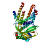
| ||||||||
|---|---|---|---|---|---|---|---|---|---|
| 1 |
| ||||||||
| Unit cell |
| ||||||||
| Components on special symmetry positions |
|
-Components
| #1: Protein | Mass: 64144.215 Da / Num. of mol.: 1 Source method: isolated from a genetically manipulated source Source: (gene. exp.) Saccharomyces cerevisiae (brewer's yeast), (gene. exp.) Influenza A virusStrain: ATCC 204508 / S288c, A/Wilson-Smith/1933 H1N1 / Gene: SMT3, YDR510W, D9719.15, PA / Production host:  Escherichia coli (E. coli) Escherichia coli (E. coli)References: UniProt: Q12306, UniProt: P15659, Hydrolases; Acting on ester bonds |
|---|---|
| #2: Chemical | ChemComp-EZS /   Mass: 232.254 Da / Num. of mol.: 1 / Source method: obtained synthetically / Formula: C13H13FN2O / Feature type: SUBJECT OF INVESTIGATION Mass: 232.254 Da / Num. of mol.: 1 / Source method: obtained synthetically / Formula: C13H13FN2O / Feature type: SUBJECT OF INVESTIGATION |
| #3: Chemical | ChemComp-DMS / Dimethyl sulfoxide  Mass: 78.133 Da / Num. of mol.: 1 / Source method: obtained synthetically / Formula: C2H6OS / Comment: DMSO, precipitant*YM Mass: 78.133 Da / Num. of mol.: 1 / Source method: obtained synthetically / Formula: C2H6OS / Comment: DMSO, precipitant*YM |
| #4: Water | ChemComp-HOH / Water Mass: 18.015 Da / Num. of mol.: 104 / Source method: isolated from a natural source / Formula: H2O Mass: 18.015 Da / Num. of mol.: 104 / Source method: isolated from a natural source / Formula: H2O |
-Experimental details
-Experiment
| Experiment | Method: X-RAY DIFFRACTION / Number of used crystals: 1 |
|---|
- Sample preparation
| Crystal | Density Matthews: 2.12 Å3/Da / Density % sol: 41.98 % |
|---|---|
| Crystal grow | Temperature: 289 K / Method: vapor diffusion, sitting drop / pH: 8.5 Details: MOLECULAR DIMENSIONS MORPHEUS SCREEN D10: 10% PEG 8000, 20% ETHYLENE GLYCOL; 20MM OF EACH 1, 6-HEXANEDIOL, 1-BUTANOL, 1,2-PROPANEDIOL, 2-PROPANOL, 1,4- BUTANEDIOL, 1,3-PROPANEDIOL; 100MM ...Details: MOLECULAR DIMENSIONS MORPHEUS SCREEN D10: 10% PEG 8000, 20% ETHYLENE GLYCOL; 20MM OF EACH 1, 6-HEXANEDIOL, 1-BUTANOL, 1,2-PROPANEDIOL, 2-PROPANOL, 1,4- BUTANEDIOL, 1,3-PROPANEDIOL; 100MM TRIS/BICINE PH 8.5; INVAA.07057.A.D15. AT 20.0 MG/ML, OVERNIGHT SOAK WITH 7 MM BSI70565, DIRECT CRYO; tray 297881d10, puck WXU6-7 PH range: 8.5 |
-Data collection
| Diffraction | Mean temperature: 100 K |
|---|---|
| Diffraction source | Source: SYNCHROTRON / Site: APS / Beamline: 21-ID-F / Wavelength: 0.97872 Å |
| Detector | Type: RAYONIX MX-300 / Detector: CCD / Date: Feb 1, 2018 |
| Radiation | Monochromator: SINGLE CRYSTAL, CYLINDRICALLY BENT, SI(220) / Protocol: SINGLE WAVELENGTH / Monochromatic (M) / Laue (L): M / Scattering type: x-ray |
| Radiation wavelength | Wavelength: 0.97872 Å / Relative weight: 1 |
| Reflection | Resolution: 2.45→50 Å / Num. obs: 21829 / % possible obs: 99.9 % / Observed criterion σ(I): -3 / Redundancy: 9.38 % / Rmerge(I) obs: 0.072 / Net I/σ(I): 21.87 |
| Reflection shell | Resolution: 2.45→2.51 Å / Rmerge(I) obs: 0.531 / Mean I/σ(I) obs: 4.72 / % possible all: 100 |
- Processing
| Software |
| ||||||||||||||||||||||||||||||||||||||||||||||||||||||||||||||||||||||||||||||||||||||||||||||||||||||||||||||||||||||||||||||||||||||||||||||||||||||
|---|---|---|---|---|---|---|---|---|---|---|---|---|---|---|---|---|---|---|---|---|---|---|---|---|---|---|---|---|---|---|---|---|---|---|---|---|---|---|---|---|---|---|---|---|---|---|---|---|---|---|---|---|---|---|---|---|---|---|---|---|---|---|---|---|---|---|---|---|---|---|---|---|---|---|---|---|---|---|---|---|---|---|---|---|---|---|---|---|---|---|---|---|---|---|---|---|---|---|---|---|---|---|---|---|---|---|---|---|---|---|---|---|---|---|---|---|---|---|---|---|---|---|---|---|---|---|---|---|---|---|---|---|---|---|---|---|---|---|---|---|---|---|---|---|---|---|---|---|---|---|---|
| Refinement | Method to determine structure: MOLECULAR REPLACEMENT Starting model: 4IUJ Resolution: 2.45→41.13 Å / SU ML: 0.27 / Cross valid method: FREE R-VALUE / Phase error: 21.19
| ||||||||||||||||||||||||||||||||||||||||||||||||||||||||||||||||||||||||||||||||||||||||||||||||||||||||||||||||||||||||||||||||||||||||||||||||||||||
| Solvent computation | Shrinkage radii: 0.9 Å / VDW probe radii: 1.11 Å | ||||||||||||||||||||||||||||||||||||||||||||||||||||||||||||||||||||||||||||||||||||||||||||||||||||||||||||||||||||||||||||||||||||||||||||||||||||||
| Refinement step | Cycle: LAST / Resolution: 2.45→41.13 Å
| ||||||||||||||||||||||||||||||||||||||||||||||||||||||||||||||||||||||||||||||||||||||||||||||||||||||||||||||||||||||||||||||||||||||||||||||||||||||
| Refine LS restraints |
| ||||||||||||||||||||||||||||||||||||||||||||||||||||||||||||||||||||||||||||||||||||||||||||||||||||||||||||||||||||||||||||||||||||||||||||||||||||||
| LS refinement shell |
| ||||||||||||||||||||||||||||||||||||||||||||||||||||||||||||||||||||||||||||||||||||||||||||||||||||||||||||||||||||||||||||||||||||||||||||||||||||||
| Refinement TLS params. | Method: refined / Refine-ID: X-RAY DIFFRACTION
| ||||||||||||||||||||||||||||||||||||||||||||||||||||||||||||||||||||||||||||||||||||||||||||||||||||||||||||||||||||||||||||||||||||||||||||||||||||||
| Refinement TLS group |
|
