 Movie
Movie Controller

[English] 日本語
 Yorodumi
- PDB-6c9u: Crystal structure of [KS3][AT3] didomain from module 3 of 6-deoxy... -
+ Open data

- Basic information
| Entry | Database: PDB / ID: 6c9u | ||||||
|---|---|---|---|---|---|---|---|
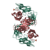
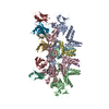
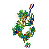
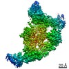
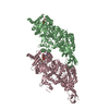
| Title | Crystal structure of [KS3][AT3] didomain from module 3 of 6-deoxyerthronolide B synthase in complex with antibody fragment (Fab) | ||||||
 Components Components |
| ||||||
 Keywords Keywords | TRANSFERASE/immune system /  DEBS / PKS / polyketide synthase / 6-deoxyerthronolide B synthase / Fab / antibody / phage display / TRANSFERASE-immune system complex DEBS / PKS / polyketide synthase / 6-deoxyerthronolide B synthase / Fab / antibody / phage display / TRANSFERASE-immune system complex | ||||||
| Function / homology |  Function and homology information Function and homology information6-deoxyerythronolide-B synthase / erythronolide synthase activity / macrolide biosynthetic process / phosphopantetheine binding / 3-oxoacyl-[acyl-carrier-protein] synthase activity / fatty acid biosynthetic process / oxidoreductase activitySimilarity search - Function | ||||||
| Biological species |  Saccharopolyspora erythraea (bacteria) Saccharopolyspora erythraea (bacteria) Homo sapiens (human) Homo sapiens (human) | ||||||
| Method | X-RAY DIFFRACTION / SYNCHROTRON / MOLECULAR REPLACEMENT / Resolution: 2.09 Å | ||||||
 Authors Authors | Deis, L.N. / Li, X. / Mathews, I.I. / Khosla, C. | ||||||
| Funding support |  United States, 1items United States, 1items
| ||||||
 Citation Citation | Journal: J. Am. Chem. Soc. / Year: 2018 Title: Structure-Function Analysis of the Extended Conformation of a Polyketide Synthase Module. Authors: Li, X. / Sevillano, N. / La Greca, F. / Deis, L. / Liu, Y.C. / Deller, M.C. / Mathews, I.I. / Matsui, T. / Cane, D.E. / Craik, C.S. / Khosla, C. | ||||||
| History |
|
- Structure visualization
| Structure viewer | Molecule: MolmilJmol/JSmol |
|---|
- Downloads & links
-Download
| PDBx/mmCIF format | 6c9u.cif.gz | 485.6 KB | Display | PDBx/mmCIF format |
|---|---|---|---|---|
| PDB format | pdb6c9u.ent.gz | 396.8 KB | Display | PDB format |
| PDBx/mmJSON format | 6c9u.json.gz | Tree view | PDBx/mmJSON format | |
| Others |  Other downloads Other downloads |
-Validation report
| Arichive directory | https://data.pdbj.org/pub/pdb/validation_reports/c9/6c9uftp://data.pdbj.org/pub/pdb/validation_reports/c9/6c9u | HTTPS FTP |
|---|
-Related structure data


-Links
 PDBj
PDBj


- Assembly
| Deposited unit | 
| ||||||||
|---|---|---|---|---|---|---|---|---|---|
| 1 | 
| ||||||||
| Unit cell |
|
-Components
-Protein , 1 types, 1 molecules A
| #1: Protein | Mass: 99797.750 Da / Num. of mol.: 1 / Fragment: [KS3][AT3] didomain from module 3 Source method: isolated from a genetically manipulated source Source: (gene. exp.) Saccharopolyspora erythraea (bacteria) / Gene: eryA / Production host: Escherichia coli (E. coli)References: UniProt: Q03132, 6-deoxyerythronolide-B synthase |
|---|
-Antibody , 2 types, 2 molecules LH
| #2: Antibody | Mass: 25715.832 Da / Num. of mol.: 1 Source method: isolated from a genetically manipulated source Source: (gene. exp.) Homo sapiens (human) / Production host: Escherichia coli (E. coli) |
|---|---|
| #3: Antibody | Mass: 26447.611 Da / Num. of mol.: 1 Source method: isolated from a genetically manipulated source Source: (gene. exp.) Homo sapiens (human) / Production host: Escherichia coli (E. coli) |
-Non-polymers , 3 types, 683 molecules 


| #4: Chemical | ChemComp-K /  Mass: 39.098 Da / Num. of mol.: 1 / Source method: obtained synthetically / Formula: K Mass: 39.098 Da / Num. of mol.: 1 / Source method: obtained synthetically / Formula: K | ||
|---|---|---|---|
| #5: Chemical |  Mass: 22.990 Da / Num. of mol.: 2 / Source method: obtained synthetically / Formula: Na Mass: 22.990 Da / Num. of mol.: 2 / Source method: obtained synthetically / Formula: Na#6: Water | ChemComp-HOH / | WaterMass: 18.015 Da / Num. of mol.: 680 / Source method: isolated from a natural source / Formula: H2O |
-Experimental details
-Experiment
| Experiment | Method: X-RAY DIFFRACTION / Number of used crystals: 1 |
|---|
- Sample preparation
| Crystal | Density Matthews: 2.85 Å3/Da / Density % sol: 56.86 % |
|---|---|
| Crystal grow | Temperature: 285 K / Method: vapor diffusion, sitting drop Details: 200 mM potassium citrate, 20%(w/v) PEG 3,350 and 12% ethylene glycol) |
-Data collection
| Diffraction | Mean temperature: 100 K |
|---|---|
| Diffraction source | Source: SYNCHROTRON / Site: SSRL / Beamline: BL12-2 / Wavelength: 1 Å |
| Detector | Type: DECTRIS PILATUS 6M-F / Detector: PIXEL / Date: Dec 17, 2015 |
| Radiation | Protocol: SINGLE WAVELENGTH / Monochromatic (M) / Laue (L): M / Scattering type: x-ray |
| Radiation wavelength | Wavelength: 1 Å / Relative weight: 1 |
| Reflection | Resolution: 2.092→46.08 Å / Num. obs: 93106 / % possible obs: 93.9 % / Redundancy: 7.4 % / Net I/σ(I): 2.44 |
| Reflection shell | Resolution: 2.09→2.17 Å |
- Processing
| Software |
| |||||||||||||||||||||||||||||||||||||||||||||||||||||||||||||||||||||||||||||||||||||||||||||||||||||||||
|---|---|---|---|---|---|---|---|---|---|---|---|---|---|---|---|---|---|---|---|---|---|---|---|---|---|---|---|---|---|---|---|---|---|---|---|---|---|---|---|---|---|---|---|---|---|---|---|---|---|---|---|---|---|---|---|---|---|---|---|---|---|---|---|---|---|---|---|---|---|---|---|---|---|---|---|---|---|---|---|---|---|---|---|---|---|---|---|---|---|---|---|---|---|---|---|---|---|---|---|---|---|---|---|---|---|---|
| Refinement | Method to determine structure: MOLECULAR REPLACEMENT Starting model: 2QO3, 1SBS Resolution: 2.09→46.08 Å / SU ML: 0.21 / Cross valid method: FREE R-VALUE / σ(F): 0 / Phase error: 21.21
| |||||||||||||||||||||||||||||||||||||||||||||||||||||||||||||||||||||||||||||||||||||||||||||||||||||||||
| Solvent computation | Shrinkage radii: 0.9 Å / VDW probe radii: 1.11 Å | |||||||||||||||||||||||||||||||||||||||||||||||||||||||||||||||||||||||||||||||||||||||||||||||||||||||||
| Refinement step | Cycle: LAST / Resolution: 2.09→46.08 Å
| |||||||||||||||||||||||||||||||||||||||||||||||||||||||||||||||||||||||||||||||||||||||||||||||||||||||||
| Refine LS restraints |
| |||||||||||||||||||||||||||||||||||||||||||||||||||||||||||||||||||||||||||||||||||||||||||||||||||||||||
| LS refinement shell |
|
