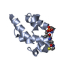
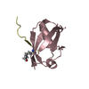
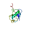
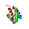
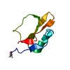
1.9 Angstrom Resolution Crystal Structure of Acyl Carrier Protein Domain (residues 1350-1461) of Polyketide Synthase Pks13 from Mycobacterium tuberculosis
Components
Polyketide synthase Pks13
Keywords
TRANSPORT PROTEIN / Structural Genomics / Center for Structural Genomics of Infectious Diseases / CSGID / Acyl Carrier Protein / Pks13
Function / homology
Function and homology information
DIM/DIP cell wall layer assembly / fatty acid synthase activity / phosphopantetheine binding / 3-oxoacyl-[acyl-carrier-protein] synthase activity / Transferases; Acyltransferases; Transferring groups other than aminoacyl groups / fatty acid biosynthetic process / cytoplasm Similarity search - Function
Method to determine structure: SAD / Resolution: 1.9→29.09 Å / Cor.coef. Fo:Fc: 0.96 / Cor.coef. Fo:Fc free: 0.949 / SU B: 5.219 / SU ML: 0.078 / Cross valid method: THROUGHOUT / ESU R: 0.13 / ESU R Free: 0.12 / Details: HYDROGENS HAVE BEEN ADDED IN THE RIDING POSITIONS
Rfactor
Num. reflection
% reflection
Selection details
Rfree
0.21731
435
5.1 %
RANDOM
Rwork
0.19346
-
-
-
obs
0.19478
8113
99.95 %
-
Solvent computation
Ion probe radii: 0.8 Å / Shrinkage radii: 0.8 Å / VDW probe radii: 1.2 Å
 Movie
Movie Controller

 Yorodumi
 Open data
 Basic information
 Components
 Keywords
 TRANSPORT PROTEIN /
  Function and homology information

 Authors
 Citation
 Structure visualization
 Downloads & links
 Other downloads
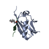


 PDBj
PDBj


 Assembly

 Mass: 65.409 Da / Num. of mol.: 2 / Source method: obtained synthetically / Formula: Zn
 Mass: 18.015 Da / Num. of mol.: 36 / Source method: isolated from a natural source / Formula: H2O
 Sample preparation
 / Beamline: 21-ID-F / Wavelength: 0.97872 Å
 Processing