 Movie
Movie Controller

[English] 日本語
 Yorodumi
- PDB-6bum: Crystal structures of cyanuric acid hydrolase from Moorella therm... -
+ Open data

- Basic information
| Entry | Database: PDB / ID: 6bum | |||||||||
|---|---|---|---|---|---|---|---|---|---|---|
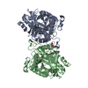
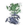
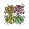
| Title | Crystal structures of cyanuric acid hydrolase from Moorella thermoacetica | |||||||||
 Components Components | Cyanuric acid amidohydrolase | |||||||||
 Keywords Keywords | HYDROLASE | |||||||||
| Function / homology |  Function and homology informationcyanuric acid amidohydrolase / cyanuric acid amidohydrolase activity / atrazine catabolic process / metal ion binding Function and homology informationcyanuric acid amidohydrolase / cyanuric acid amidohydrolase activity / atrazine catabolic process / metal ion bindingSimilarity search - Function | |||||||||
| Biological species |   Moorella thermoacetica (bacteria) Moorella thermoacetica (bacteria) | |||||||||
| Method | X-RAY DIFFRACTION / SYNCHROTRON / MOLECULAR REPLACEMENT / Resolution: 1.51 Å | |||||||||
 Authors Authors | Shi, K. / Cho, S. / Seffernick, J.L. / Bera, A. / Wackett, L.P. / Aihara, H. | |||||||||
| Funding support |  United States, 2items United States, 2items
| |||||||||
 Citation Citation | Journal: Plos One / Year: 2019 Title: Crystal structures of Moorella thermoacetica cyanuric acid hydrolase reveal conformational flexibility and asymmetry important for catalysis. Authors: Shi, K. / Cho, S. / Aukema, K.G. / Lee, T. / Bera, A.K. / Seffernick, J.L. / Wackett, L.P. / Aihara, H. | |||||||||
| History |
|
- Structure visualization
| Structure viewer | Molecule: MolmilJmol/JSmol |
|---|
- Downloads & links
-Download
| PDBx/mmCIF format | 6bum.cif.gz | 808 KB | Display | PDBx/mmCIF format |
|---|---|---|---|---|
| PDB format | pdb6bum.ent.gz | 683.5 KB | Display | PDB format |
| PDBx/mmJSON format | 6bum.json.gz | Tree view | PDBx/mmJSON format | |
| Others |  Other downloads Other downloads |
-Validation report
| Arichive directory | https://data.pdbj.org/pub/pdb/validation_reports/bu/6bumftp://data.pdbj.org/pub/pdb/validation_reports/bu/6bum | HTTPS FTP |
|---|
-Related structure data
| Related structure data |  6bunC  6buoC  6bupC  6buqC  6burC  6cwjC  6dhjC  3nq4S S: Starting model for refinement C: citing same article ( |
|---|---|
| Similar structure data |


-Links
 PDBj
PDBj- Assembly
| Deposited unit | 
| ||||||||
|---|---|---|---|---|---|---|---|---|---|
| 1 |
| ||||||||
| Unit cell |
|
-Components
-Protein , 1 types, 4 molecules ABCD
| #1: Protein | / CAH / Barbiturase Mass: 38484.652 Da / Num. of mol.: 4 Mutation: Q102A, E102A, K107A, L279I, K280R, F281S, E288D, L290M, A291D, K292R Source method: isolated from a genetically manipulated source Source: (gene. exp.) Moorella thermoacetica (strain ATCC 39073 / JCM 9320) (bacteria)Strain: ATCC 39073 / JCM 9320 / Gene: Moth_2120 / Production host: Escherichia coli (E. coli) / References: UniProt: Q2RGM7, cyanuric acid amidohydrolase |
|---|
-Non-polymers , 5 types, 1200 molecules 


| #2: Chemical | ChemComp-MLI / Malonic acid Mass: 102.046 Da / Num. of mol.: 4 / Source method: obtained synthetically / Formula: C3H2O4 Mass: 102.046 Da / Num. of mol.: 4 / Source method: obtained synthetically / Formula: C3H2O4#3: Chemical | ChemComp-CA /  Mass: 40.078 Da / Num. of mol.: 4 / Source method: obtained synthetically / Formula: Ca Mass: 40.078 Da / Num. of mol.: 4 / Source method: obtained synthetically / Formula: Ca#4: Chemical | ChemComp-PDO / 1,3-Propanediol Mass: 76.094 Da / Num. of mol.: 24 / Source method: obtained synthetically / Formula: C3H8O2 Mass: 76.094 Da / Num. of mol.: 24 / Source method: obtained synthetically / Formula: C3H8O2#5: Chemical | ChemComp-CL / | Chloride Mass: 35.453 Da / Num. of mol.: 1 / Source method: obtained synthetically / Formula: Cl Mass: 35.453 Da / Num. of mol.: 1 / Source method: obtained synthetically / Formula: Cl#6: Water | ChemComp-HOH / | WaterMass: 18.015 Da / Num. of mol.: 1167 / Source method: isolated from a natural source / Formula: H2O |
|---|
-Experimental details
-Experiment
| Experiment | Method: X-RAY DIFFRACTION / Number of used crystals: 1 |
|---|
- Sample preparation
| Crystal | Density Matthews: 2.4 Å3/Da / Density % sol: 48.67 % |
|---|---|
| Crystal grow | Temperature: 293 K / Method: vapor diffusion, sitting drop / pH: 5 / Details: 20%PEG3350, 100mMCaCl2 |
-Data collection
| Diffraction | Mean temperature: 100 K |
|---|---|
| Diffraction source | Source: SYNCHROTRON / Site: APS / Beamline: 24-ID-E / Wavelength: 0.979 Å |
| Detector | Type: DECTRIS PILATUS3 S 6M / Detector: PIXEL / Date: Dec 6, 2015 |
| Radiation | Protocol: SINGLE WAVELENGTH / Monochromatic (M) / Laue (L): M / Scattering type: x-ray |
| Radiation wavelength | Wavelength: 0.979 Å / Relative weight: 1 |
| Reflection | Resolution: 1.51→44.35 Å / Num. obs: 221630 / % possible obs: 95.8 % / Redundancy: 3.9 % / CC1/2: 0.999 / Rmerge(I) obs: 0.037 / Rsym value: 0.037 / Net I/σ(I): 20.1 |
| Reflection shell | Resolution: 1.51→1.54 Å / Redundancy: 2 % / Rmerge(I) obs: 0.625 / Mean I/σ(I) obs: 1.2 / Num. unique obs: 8020 / CC1/2: 0.471 / % possible all: 70.2 |
- Processing
| Software |
| |||||||||||||||||||||||||||||||||||||||||||||||||||||||||||||||||||||||||||||||||||||||||||||||||||||||||||||||||||||||||||||||||||||||||||||||||||||||||||||||||||||||||||||||||||||||||||||||||||||||||||||||||||||||||
|---|---|---|---|---|---|---|---|---|---|---|---|---|---|---|---|---|---|---|---|---|---|---|---|---|---|---|---|---|---|---|---|---|---|---|---|---|---|---|---|---|---|---|---|---|---|---|---|---|---|---|---|---|---|---|---|---|---|---|---|---|---|---|---|---|---|---|---|---|---|---|---|---|---|---|---|---|---|---|---|---|---|---|---|---|---|---|---|---|---|---|---|---|---|---|---|---|---|---|---|---|---|---|---|---|---|---|---|---|---|---|---|---|---|---|---|---|---|---|---|---|---|---|---|---|---|---|---|---|---|---|---|---|---|---|---|---|---|---|---|---|---|---|---|---|---|---|---|---|---|---|---|---|---|---|---|---|---|---|---|---|---|---|---|---|---|---|---|---|---|---|---|---|---|---|---|---|---|---|---|---|---|---|---|---|---|---|---|---|---|---|---|---|---|---|---|---|---|---|---|---|---|---|---|---|---|---|---|---|---|---|---|---|---|---|---|---|---|---|
| Refinement | Method to determine structure: MOLECULAR REPLACEMENT Starting model: 3NQ4 Resolution: 1.51→29.993 Å / SU ML: 0.12 / Cross valid method: FREE R-VALUE / σ(F): 1.33 / Phase error: 13.91
| |||||||||||||||||||||||||||||||||||||||||||||||||||||||||||||||||||||||||||||||||||||||||||||||||||||||||||||||||||||||||||||||||||||||||||||||||||||||||||||||||||||||||||||||||||||||||||||||||||||||||||||||||||||||||
| Solvent computation | Shrinkage radii: 0.9 Å / VDW probe radii: 1.11 Å | |||||||||||||||||||||||||||||||||||||||||||||||||||||||||||||||||||||||||||||||||||||||||||||||||||||||||||||||||||||||||||||||||||||||||||||||||||||||||||||||||||||||||||||||||||||||||||||||||||||||||||||||||||||||||
| Refinement step | Cycle: LAST / Resolution: 1.51→29.993 Å
| |||||||||||||||||||||||||||||||||||||||||||||||||||||||||||||||||||||||||||||||||||||||||||||||||||||||||||||||||||||||||||||||||||||||||||||||||||||||||||||||||||||||||||||||||||||||||||||||||||||||||||||||||||||||||
| Refine LS restraints |
| |||||||||||||||||||||||||||||||||||||||||||||||||||||||||||||||||||||||||||||||||||||||||||||||||||||||||||||||||||||||||||||||||||||||||||||||||||||||||||||||||||||||||||||||||||||||||||||||||||||||||||||||||||||||||
| LS refinement shell |
|
