[English] 日本語
Yorodumi
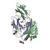
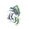
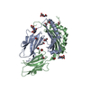
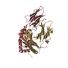
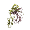
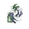
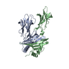
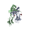
- PDB-6blr: Crystal Structure of IAg7 in complex with insulin mimotope p8E9E6SS -

+
Open data


ID or keywords:

Loading...

-
Basic information

Entry
Database: PDB / ID: 6blr
TitleCrystal Structure of IAg7 in complex with insulin mimotope p8E9E6SS
Components
  • H2-Ab1 protein
  • IAg7 alpha chain
KeywordsIMMUNE SYSTEM / INSULIN / TYPE I DIABETES / T CELL / AUTOIMMUNE DISEASE
Function / homology
Function and homology information


antigen processing and presentation of peptide antigen / positive regulation of T cell differentiation / antigen processing and presentation / negative regulation of T cell proliferation / multivesicular body / peptide antigen assembly with MHC class II protein complex / MHC class II protein complex / peptide antigen binding / antigen processing and presentation of exogenous peptide antigen via MHC class II / positive regulation of immune response ...antigen processing and presentation of peptide antigen / positive regulation of T cell differentiation / antigen processing and presentation / negative regulation of T cell proliferation / multivesicular body / peptide antigen assembly with MHC class II protein complex / MHC class II protein complex / peptide antigen binding / antigen processing and presentation of exogenous peptide antigen via MHC class II / positive regulation of immune response / positive regulation of T cell activation / MHC class II protein complex binding / late endosome membrane / adaptive immune response / lysosome / early endosome / lysosomal membrane / external side of plasma membrane / protein-containing complex binding / Golgi apparatus / cell surface / plasma membrane
Similarity search - Function
Class II Histocompatibility Antigen, M Beta Chain; Chain B, domain 1 / Class II Histocompatibility Antigen, M Beta Chain; Chain B, domain 1 / MHC class II, beta chain, N-terminal / Class II histocompatibility antigen, beta domain / Class II histocompatibility antigen, beta domain / MHC class II, alpha chain, N-terminal / Class II histocompatibility antigen, alpha domain / Class II histocompatibility antigen, alpha domain / MHC class II, alpha/beta chain, N-terminal / MHC classes I/II-like antigen recognition protein ...Class II Histocompatibility Antigen, M Beta Chain; Chain B, domain 1 / Class II Histocompatibility Antigen, M Beta Chain; Chain B, domain 1 / MHC class II, beta chain, N-terminal / Class II histocompatibility antigen, beta domain / Class II histocompatibility antigen, beta domain / MHC class II, alpha chain, N-terminal / Class II histocompatibility antigen, alpha domain / Class II histocompatibility antigen, alpha domain / MHC class II, alpha/beta chain, N-terminal / MHC classes I/II-like antigen recognition protein / Immunoglobulin/major histocompatibility complex, conserved site / Immunoglobulins and major histocompatibility complex proteins signature. / Immunoglobulin C-Type / Immunoglobulin C1-set / Immunoglobulin C1-set domain / Ig-like domain profile. / Immunoglobulin-like domain / Immunoglobulin-like domain superfamily / Immunoglobulins / Roll / Immunoglobulin-like fold / Immunoglobulin-like / Sandwich / Mainly Beta / Alpha Beta
Similarity search - Domain/homology
H-2 class II histocompatibility antigen, A-D alpha chain / H2-Ab1 protein
Similarity search - Component
Biological speciesMus musculus (house mouse)
MethodX-RAY DIFFRACTION / SYNCHROTRON / Resolution: 1.96 Å
AuthorsWang, Y. / Dai, S.
Funding support United States, 2items
OrganizationGrant numberCountry
National Institutes of Health/National Institute of Environmental Health Sciences (NIH/NIEHS)ES-025797 United States
National Institutes of Health/National Institute Of Allergy and Infectious Diseases (NIH/NIAID)5T32AI074491-08 United States
CitationJournal: Proc. Natl. Acad. Sci. U.S.A. / Year: 2018
Title: C-terminal modification of the insulin B:11-23 peptide creates superagonists in mouse and human type 1 diabetes.
Authors: Wang, Y. / Sosinowski, T. / Novikov, A. / Crawford, F. / Neau, D.B. / Yang, J. / Kwok, W.W. / Marrack, P. / Kappler, J.W. / Dai, S.
History
DepositionNov 11, 2017Deposition site: RCSB / Processing site: RCSB
Revision 1.0Dec 20, 2017Provider: repository / Type: Initial release
Revision 1.1Jan 3, 2018Group: Database references / Category: citation / citation_author
Item: _citation.country / _citation.journal_abbrev ..._citation.country / _citation.journal_abbrev / _citation.journal_id_ASTM / _citation.journal_id_CSD / _citation.journal_id_ISSN / _citation.pdbx_database_id_DOI / _citation.pdbx_database_id_PubMed / _citation.title / _citation.year
Revision 1.2Jan 10, 2018Group: Database references / Category: citation
Item: _citation.journal_volume / _citation.page_first ..._citation.journal_volume / _citation.page_first / _citation.page_last / _citation.year
Revision 1.3Jan 17, 2018Group: Author supporting evidence / Category: pdbx_audit_support / Item: _pdbx_audit_support.funding_organization
Revision 1.4Mar 14, 2018Group: Database references / Category: struct_ref_seq_dif / Item: _struct_ref_seq_dif.details
Revision 1.5Dec 11, 2019Group: Author supporting evidence / Database references / Category: pdbx_audit_support / struct_ref_seq_dif
Item: _pdbx_audit_support.funding_organization / _struct_ref_seq_dif.details

-
Structure visualization

Structure viewerMolecule:
MolmilJmol/JSmol

Downloads & links

-
Assembly

Deposited unit
A: IAg7 alpha chain
B: H2-Ab1 protein
hetero molecules


Theoretical massNumber of molelcules
Total (without water)46,3213
Polymers46,2592
Non-polymers621
Water6,125340
1


  • Idetical with deposited unit
  • defined by author&software
  • Evidence: gel filtration
TypeNameSymmetry operationNumber
identity operation1_555x,y,z1
Buried area6710 Å2
ΔGint-28 kcal/mol
Surface area17930 Å2
MethodPISA
Unit cell
Length a, b, c (Å)89.736, 111.379, 95.275
Angle α, β, γ (deg.)90.00, 90.00, 90.00
Int Tables number20
Space group name H-MC2221

-
Components

#1: Protein IAg7 alpha chain


Mass: 20896.387 Da / Num. of mol.: 1 / Mutation: N64C
Source method: isolated from a genetically manipulated source
Source: (gene. exp.) Mus musculus (house mouse) / Cell line (production host): Hi5 / Production host: Trichoplusia ni (cabbage looper) / References: UniProt: P04228
#2: Protein H2-Ab1 protein


Mass: 25362.342 Da / Num. of mol.: 1
Source method: isolated from a genetically manipulated source
Source: (gene. exp.) Mus musculus (house mouse) / Gene: H2-Ab1 / Cell line (production host): Hi5 / Production host: Trichoplusia ni (cabbage looper) / References: UniProt: Q31135
#3: Chemical ChemComp-EDO / 1,2-ETHANEDIOL / ETHYLENE GLYCOL / Ethylene glycol


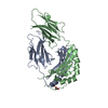
Mass: 62.068 Da / Num. of mol.: 1 / Source method: obtained synthetically / Formula: C2H6O2
#4: Water ChemComp-HOH / water / Water


Mass: 18.015 Da / Num. of mol.: 340 / Source method: isolated from a natural source / Formula: H2O
Sequence detailsinsulin mimotope

-
Experimental details

-
Experiment

ExperimentMethod: X-RAY DIFFRACTION / Number of used crystals: 1

-
Sample preparation

CrystalDensity Matthews: 2.57 Å3/Da / Density % sol: 52.2 %
Crystal growTemperature: 278 K / Method: vapor diffusion, hanging drop
Details: 15% PEG4000, 10mM sodium citrate at pH5.0, 100 mM MgCl2

-
Data collection

DiffractionMean temperature: 100 K
Diffraction sourceSource: SYNCHROTRON / Site: APS / Beamline: 24-ID-C / Wavelength: 0.9795 Å
DetectorType: ADSC QUANTUM 315 / Detector: CCD / Date: Jan 24, 2014
RadiationProtocol: SINGLE WAVELENGTH / Monochromatic (M) / Laue (L): M / Scattering type: x-ray
Radiation wavelengthWavelength: 0.9795 Å / Relative weight: 1
ReflectionResolution: 1.96→50 Å / Num. obs: 24967 / % possible obs: 74.6 % / Redundancy: 4.5 % / Rmerge(I) obs: 0.09 / Net I/σ(I): 11.7
Reflection shellResolution: 1.96→2.04 Å

-
Processing

Software
NameVersionClassification
PHENIX(1.12_2829: ???)refinement
HKL-2000data reduction
HKL-2000data scaling
PHASERphasing
RefinementResolution: 1.96→40.592 Å / SU ML: 0.2 / Cross valid method: FREE R-VALUE / σ(F): 1.36 / Phase error: 22.15
RfactorNum. reflection% reflection
Rfree0.2277 1229 4.92 %
Rwork0.1706 --
obs0.1734 24967 72.09 %
Solvent computationShrinkage radii: 0.9 Å / VDW probe radii: 1.11 Å
Refinement stepCycle: LAST / Resolution: 1.96→40.592 Å
ProteinNucleic acidLigandSolventTotal
Num. atoms3072 0 4 340 3416
Refine LS restraints
Refine-IDTypeDev idealNumber
X-RAY DIFFRACTIONf_bond_d0.0073188
X-RAY DIFFRACTIONf_angle_d0.9744340
X-RAY DIFFRACTIONf_dihedral_angle_d7.3452236
X-RAY DIFFRACTIONf_chiral_restr0.062469
X-RAY DIFFRACTIONf_plane_restr0.005563
LS refinement shell
Resolution (Å)Rfactor RfreeNum. reflection RfreeRfactor RworkNum. reflection RworkRefine-ID% reflection obs (%)
1.9594-2.03780.27241040.1862114X-RAY DIFFRACTION58
2.0378-2.13060.2491280.18392714X-RAY DIFFRACTION75
2.1306-2.24290.2551420.17672798X-RAY DIFFRACTION77
2.2429-2.38340.24451460.18192786X-RAY DIFFRACTION77
2.3834-2.56740.2681380.18482748X-RAY DIFFRACTION76
2.5674-2.82570.25311560.1862709X-RAY DIFFRACTION75
2.8257-3.23440.24511410.18082716X-RAY DIFFRACTION74
3.2344-4.07440.18871480.15712596X-RAY DIFFRACTION71
4.0744-40.60070.20461260.15312557X-RAY DIFFRACTION67

+
About Yorodumi

-
News

-
Feb 9, 2022. New format data for meta-information of EMDB entries

New format data for meta-information of EMDB entries

  • Version 3 of the EMDB header file is now the official format.
  • The previous official version 1.9 will be removed from the archive.

Related info.:EMDB header

External links:wwPDB to switch to version 3 of the EMDB data model

-
Aug 12, 2020. Covid-19 info

Covid-19 info

URL: https://pdbj.org/emnavi/covid19.php

New page: Covid-19 featured information page in EM Navigator.

Related info.:Covid-19 info / Mar 5, 2020. Novel coronavirus structure data

+
Mar 5, 2020. Novel coronavirus structure data

Novel coronavirus structure data

Related info.:Yorodumi Speices / Aug 12, 2020. Covid-19 info

External links:COVID-19 featured content - PDBj / Molecule of the Month (242):Coronavirus Proteases

+
Jan 31, 2019. EMDB accession codes are about to change! (news from PDBe EMDB page)

EMDB accession codes are about to change! (news from PDBe EMDB page)

  • The allocation of 4 digits for EMDB accession codes will soon come to an end. Whilst these codes will remain in use, new EMDB accession codes will include an additional digit and will expand incrementally as the available range of codes is exhausted. The current 4-digit format prefixed with “EMD-” (i.e. EMD-XXXX) will advance to a 5-digit format (i.e. EMD-XXXXX), and so on. It is currently estimated that the 4-digit codes will be depleted around Spring 2019, at which point the 5-digit format will come into force.
  • The EM Navigator/Yorodumi systems omit the EMD- prefix.

Related info.:Q: What is EMD? / ID/Accession-code notation in Yorodumi/EM Navigator

External links:EMDB Accession Codes are Changing Soon! / Contact to PDBj

+
Jul 12, 2017. Major update of PDB

Major update of PDB

  • wwPDB released updated PDB data conforming to the new PDBx/mmCIF dictionary.
  • This is a major update changing the version number from 4 to 5, and with Remediation, in which all the entries are updated.
  • In this update, many items about electron microscopy experimental information are reorganized (e.g. em_software).
  • Now, EM Navigator and Yorodumi are based on the updated data.

External links:wwPDB Remediation / Enriched Model Files Conforming to OneDep Data Standards Now Available in the PDB FTP Archive

-
Yorodumi

Thousand views of thousand structures

  • Yorodumi is a browser for structure data from EMDB, PDB, SASBDB, etc.
  • This page is also the successor to EM Navigator detail page, and also detail information page/front-end page for Omokage search.
  • The word "yorodu" (or yorozu) is an old Japanese word meaning "ten thousand". "mi" (miru) is to see.

Related info.:EMDB / PDB / SASBDB / Comparison of 3 databanks / Yorodumi Search / Aug 31, 2016. New EM Navigator & Yorodumi / Yorodumi Papers / Jmol/JSmol / Function and homology information / Changes in new EM Navigator and Yorodumi

Read more