 Movie
Movie Controller


+ Open data

- Basic information
| Entry | Database: PDB / ID: 6bhx | ||||||
|---|---|---|---|---|---|---|---|
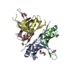
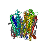
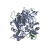
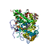
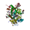
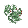
| Title | B. subtilis SsbA with DNA | ||||||
 Components Components |
| ||||||
 Keywords Keywords | DNA binding protein/DNA /  Single-stranded DNA binding protein / DNA Replication / DNA Repair / DNA binding protein-DNA complex Single-stranded DNA binding protein / DNA Replication / DNA Repair / DNA binding protein-DNA complex | ||||||
| Function / homology |  Function and homology information Function and homology informationpositive regulation of helicase activity / nucleoid / single-stranded DNA binding / DNA recombination / DNA replication / DNA repairSimilarity search - Function | ||||||
| Biological species |  Bacillus subtilis (bacteria) Bacillus subtilis (bacteria)synthetic construct (others) | ||||||
| Method | X-RAY DIFFRACTION / SYNCHROTRON / MOLECULAR REPLACEMENT / Resolution: 2.936 Å | ||||||
 Authors Authors | Dubiel, K.D. / Myers, A.R. / Satyshur, K.A. / Keck, J.L. | ||||||
| Funding support |  United States, 1items United States, 1items
| ||||||
 Citation Citation | Journal: J. Mol. Biol. / Year: 2019 Title: Structural Mechanisms of Cooperative DNA Binding by Bacterial Single-Stranded DNA-Binding Proteins. Authors: Dubiel, K. / Myers, A.R. / Kozlov, A.G. / Yang, O. / Zhang, J. / Ha, T. / Lohman, T.M. / Keck, J.L. | ||||||
| History |
|
- Structure visualization
| Structure viewer | Molecule: MolmilJmol/JSmol |
|---|
- Downloads & links
-Download
| PDBx/mmCIF format | 6bhx.cif.gz | 163.5 KB | Display | PDBx/mmCIF format |
|---|---|---|---|---|
| PDB format | pdb6bhx.ent.gz | 129 KB | Display | PDB format |
| PDBx/mmJSON format | 6bhx.json.gz | Tree view | PDBx/mmJSON format | |
| Others |  Other downloads Other downloads |
-Validation report
| Arichive directory | https://data.pdbj.org/pub/pdb/validation_reports/bh/6bhxftp://data.pdbj.org/pub/pdb/validation_reports/bh/6bhx | HTTPS FTP |
|---|
-Related structure data


-Links
 PDBj
PDBj


- Assembly
| Deposited unit | 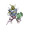
| ||||||||
|---|---|---|---|---|---|---|---|---|---|
| 1 |
| ||||||||
| Unit cell |
|
-Components
| #1: Protein | Mass: 14592.171 Da / Num. of mol.: 4 Source method: isolated from a genetically manipulated source Source: (gene. exp.) Bacillus subtilis (strain 168) (bacteria)Strain: 168 / Gene: ssbA, BSU40900 / Plasmid: pET11a / Production host: Escherichia coli (E. coli) / Strain (production host): BL21 (DE3) / References: UniProt: P37455#2: DNA chain | | Mass: 9080.827 Da / Num. of mol.: 1 / Source method: obtained synthetically / Source: (synth.) synthetic construct (others) #3: Water | ChemComp-HOH / | Water Mass: 18.015 Da / Num. of mol.: 31 / Source method: isolated from a natural source / Formula: H2O Mass: 18.015 Da / Num. of mol.: 31 / Source method: isolated from a natural source / Formula: H2O |
|---|
-Experimental details
-Experiment
| Experiment | Method: X-RAY DIFFRACTION / Number of used crystals: 1 |
|---|
- Sample preparation
| Crystal | Density Matthews: 2.03 Å3/Da / Density % sol: 39.45 % |
|---|---|
| Crystal grow | Temperature: 293 K / Method: vapor diffusion, hanging drop Details: 50% mixture with 50 mM MES pH 6.5, 7% PEG 8000, 100 mM magnesium acetate, 200 mM potassium chloride SsbA was incubated with a 1:2 SsbA to dT35 ratio and a-chymotrypsin prior to crystallization PH range: 6.5 - 8 |
-Data collection
| Diffraction | Mean temperature: 100 K |
|---|---|
| Diffraction source | Source: SYNCHROTRON / Site: APS / Beamline: 21-ID-F / Wavelength: 0.97872 Å |
| Detector | Type: MARMOSAIC 225 mm CCD / Detector: CCD / Date: Mar 23, 2013 |
| Radiation | Protocol: SINGLE WAVELENGTH / Monochromatic (M) / Laue (L): M / Scattering type: x-ray |
| Radiation wavelength | Wavelength: 0.97872 Å / Relative weight: 1 |
| Reflection | Resolution: 2.92→50 Å / Num. obs: 12270 / % possible obs: 99.7 % / Redundancy: 7.6 % / Rmerge(I) obs: 0.128 / Net I/σ(I): 14.48 |
| Reflection shell | Resolution: 2.92→2.97 Å / Redundancy: 6.3 % / Rmerge(I) obs: 0.496 / Mean I/σ(I) obs: 2.29 / Num. unique obs: 597 / % possible all: 98.4 |
- Processing
| Software |
| |||||||||||||||||||||||||||||||||||
|---|---|---|---|---|---|---|---|---|---|---|---|---|---|---|---|---|---|---|---|---|---|---|---|---|---|---|---|---|---|---|---|---|---|---|---|---|
| Refinement | Method to determine structure: MOLECULAR REPLACEMENT / Resolution: 2.936→44.284 Å / SU ML: 0.43 / Cross valid method: FREE R-VALUE / σ(F): 1.35 / Phase error: 26.22
| |||||||||||||||||||||||||||||||||||
| Solvent computation | Shrinkage radii: 0.9 Å / VDW probe radii: 1.11 Å | |||||||||||||||||||||||||||||||||||
| Refinement step | Cycle: LAST / Resolution: 2.936→44.284 Å
| |||||||||||||||||||||||||||||||||||
| Refine LS restraints |
| |||||||||||||||||||||||||||||||||||
| LS refinement shell |
|
