 Movie
Movie Controller

[English] 日本語
 Yorodumi
- PDB-6axt: Structure of the T58S/T107I/P122A mutant of the HIV-1 capsid protein -
+ Open data

- Basic information
| Entry | Database: PDB / ID: 6axt | ||||||
|---|---|---|---|---|---|---|---|
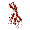
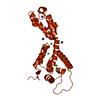
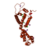
| Title | Structure of the T58S/T107I/P122A mutant of the HIV-1 capsid protein | ||||||
 Components Components | HIV-1 capsid protein | ||||||
 Keywords Keywords |  VIRAL PROTEIN / HIV-1 capsid protein / hexamer / T58S/T107I/P122A mutant VIRAL PROTEIN / HIV-1 capsid protein / hexamer / T58S/T107I/P122A mutant | ||||||
| Function / homology |  Function and homology information Function and homology informationviral budding via host ESCRT complex / viral process / ISG15 antiviral mechanism / host multivesicular body / viral nucleocapsid / host cell cytoplasm / host cell nucleus / host cell plasma membrane / virion membrane / structural molecule activity ...viral budding via host ESCRT complex / viral process / ISG15 antiviral mechanism / host multivesicular body / viral nucleocapsid / host cell cytoplasm / host cell nucleus / host cell plasma membrane / virion membrane / structural molecule activity / RNA binding / zinc ion binding / membrane / cytoplasmSimilarity search - Function | ||||||
| Biological species |   Human immunodeficiency virus 1 Human immunodeficiency virus 1 | ||||||
| Method | X-RAY DIFFRACTION / SYNCHROTRON / MOLECULAR REPLACEMENT / molecular replacement / Resolution: 2.4 Å | ||||||
 Authors Authors | Gres, A.T. / Kirby, K.A. / Sarafianos, S.G. | ||||||
| Funding support |  United States, 1items United States, 1items
| ||||||
 Citation Citation | Journal: To be published Title: Identification of a novel element in HIV-1 capsid critical for assembly and maturation Authors: Novikova, M. / Gres, A.T. / Kirby, K.A. / Sarafianos, S.G. / Freed, E.O. | ||||||
| History |
|
- Structure visualization
| Structure viewer | Molecule: MolmilJmol/JSmol |
|---|
- Downloads & links
-Download
| PDBx/mmCIF format | 6axt.cif.gz | 103.7 KB | Display | PDBx/mmCIF format |
|---|---|---|---|---|
| PDB format | pdb6axt.ent.gz | 78.2 KB | Display | PDB format |
| PDBx/mmJSON format | 6axt.json.gz | Tree view | PDBx/mmJSON format | |
| Others |  Other downloads Other downloads |
-Validation report
| Arichive directory | https://data.pdbj.org/pub/pdb/validation_reports/ax/6axtftp://data.pdbj.org/pub/pdb/validation_reports/ax/6axt | HTTPS FTP |
|---|
-Related structure data
| Related structure data |  6axsC  6axvC  6axxC  6axyC  4xfxS S: Starting model for refinement C: citing same article ( |
|---|---|
| Similar structure data |


-Links
 PDBj
PDBj

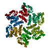
- Assembly
| Deposited unit | 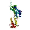
| ||||||||
|---|---|---|---|---|---|---|---|---|---|
| 1 | x 6
| ||||||||
| Unit cell |
|
-Components
| #1: Protein | Mass: 25602.416 Da / Num. of mol.: 1 / Fragment: UNP residues 133-363 / Mutation: T58S, T107I, P122A Source method: isolated from a genetically manipulated source Source: (gene. exp.) Human immunodeficiency virus 1 / Gene: gag / Plasmid: pET11a / Production host:  Escherichia coli (E. coli) / Strain (production host): BL21(DE3) / References: UniProt: B6DRA0, UniProt: P12493*PLUS Escherichia coli (E. coli) / Strain (production host): BL21(DE3) / References: UniProt: B6DRA0, UniProt: P12493*PLUS | ||||
|---|---|---|---|---|---|
| #2: Chemical | ChemComp-IOD / Iodide  Mass: 126.904 Da / Num. of mol.: 9 / Source method: obtained synthetically / Formula: I Mass: 126.904 Da / Num. of mol.: 9 / Source method: obtained synthetically / Formula: I#3: Chemical | ChemComp-CL / Chloride  Mass: 35.453 Da / Num. of mol.: 6 / Source method: obtained synthetically / Formula: Cl Mass: 35.453 Da / Num. of mol.: 6 / Source method: obtained synthetically / Formula: Cl#4: Water | ChemComp-HOH / | Water Mass: 18.015 Da / Num. of mol.: 41 / Source method: isolated from a natural source / Formula: H2O Mass: 18.015 Da / Num. of mol.: 41 / Source method: isolated from a natural source / Formula: H2O |
-Experimental details
-Experiment
| Experiment | Method: X-RAY DIFFRACTION / Number of used crystals: 1 |
|---|
- Sample preparation
| Crystal | Density Matthews: 2.77 Å3/Da / Density % sol: 55.57 % / Mosaicity: 0.21 ° |
|---|---|
| Crystal grow | Temperature: 291 K / Method: vapor diffusion, hanging drop / Details: PEG3350, NaI, MIB, Glycerol |
-Data collection
| Diffraction | Mean temperature: 100 K | ||||||||||||||||||||||||
|---|---|---|---|---|---|---|---|---|---|---|---|---|---|---|---|---|---|---|---|---|---|---|---|---|---|
| Diffraction source | Source: SYNCHROTRON / Site: APS / Beamline: 23-ID-B / Wavelength: 1.03376 Å | ||||||||||||||||||||||||
| Detector | Type: MARMOSAIC 300 mm CCD / Detector: CCD / Date: Nov 8, 2015 | ||||||||||||||||||||||||
| Radiation | Protocol: SINGLE WAVELENGTH / Monochromatic (M) / Laue (L): M / Scattering type: x-ray | ||||||||||||||||||||||||
| Radiation wavelength | Wavelength: 1.03376 Å / Relative weight: 1 | ||||||||||||||||||||||||
| Reflection | Resolution: 2.4→46.7 Å / Num. obs: 10448 / % possible obs: 94.1 % / Redundancy: 8.3 % / Biso Wilson estimate: 60.44 Å2 / CC1/2: 0.999 / Rmerge(I) obs: 0.071 / Rpim(I) all: 0.026 / Rrim(I) all: 0.075 / Net I/σ(I): 17.7 / Num. measured all: 86620 / Scaling rejects: 0 | ||||||||||||||||||||||||
| Reflection shell | Diffraction-ID: 1
|
-Phasing
| Phasing | Method: molecular replacement | |||||||||
|---|---|---|---|---|---|---|---|---|---|---|
| Phasing MR | Model details: Phaser MODE: MR_AUTO
|
- Processing
| Software |
| ||||||||||||||||||||||||||||||||||||||||||||||||||||||||||||||||||||||||||||||||||||||||||||||||||||||||||||||||||||||||||||||||||||||||||||||||||||||||||||||||||||||||||||||||||||||||||||||||||||||||
|---|---|---|---|---|---|---|---|---|---|---|---|---|---|---|---|---|---|---|---|---|---|---|---|---|---|---|---|---|---|---|---|---|---|---|---|---|---|---|---|---|---|---|---|---|---|---|---|---|---|---|---|---|---|---|---|---|---|---|---|---|---|---|---|---|---|---|---|---|---|---|---|---|---|---|---|---|---|---|---|---|---|---|---|---|---|---|---|---|---|---|---|---|---|---|---|---|---|---|---|---|---|---|---|---|---|---|---|---|---|---|---|---|---|---|---|---|---|---|---|---|---|---|---|---|---|---|---|---|---|---|---|---|---|---|---|---|---|---|---|---|---|---|---|---|---|---|---|---|---|---|---|---|---|---|---|---|---|---|---|---|---|---|---|---|---|---|---|---|---|---|---|---|---|---|---|---|---|---|---|---|---|---|---|---|---|---|---|---|---|---|---|---|---|---|---|---|---|---|---|---|---|
| Refinement | Method to determine structure: MOLECULAR REPLACEMENT Starting model: 4XFX Resolution: 2.4→46.7 Å / SU ML: 0.3 / Cross valid method: FREE R-VALUE / σ(F): 1.35 / Phase error: 31.55
| ||||||||||||||||||||||||||||||||||||||||||||||||||||||||||||||||||||||||||||||||||||||||||||||||||||||||||||||||||||||||||||||||||||||||||||||||||||||||||||||||||||||||||||||||||||||||||||||||||||||||
| Solvent computation | Shrinkage radii: 0.9 Å / VDW probe radii: 1.11 Å | ||||||||||||||||||||||||||||||||||||||||||||||||||||||||||||||||||||||||||||||||||||||||||||||||||||||||||||||||||||||||||||||||||||||||||||||||||||||||||||||||||||||||||||||||||||||||||||||||||||||||
| Displacement parameters | Biso max: 174.51 Å2 / Biso mean: 79.5318 Å2 / Biso min: 37.8 Å2 | ||||||||||||||||||||||||||||||||||||||||||||||||||||||||||||||||||||||||||||||||||||||||||||||||||||||||||||||||||||||||||||||||||||||||||||||||||||||||||||||||||||||||||||||||||||||||||||||||||||||||
| Refinement step | Cycle: final / Resolution: 2.4→46.7 Å
| ||||||||||||||||||||||||||||||||||||||||||||||||||||||||||||||||||||||||||||||||||||||||||||||||||||||||||||||||||||||||||||||||||||||||||||||||||||||||||||||||||||||||||||||||||||||||||||||||||||||||
| Refine LS restraints |
| ||||||||||||||||||||||||||||||||||||||||||||||||||||||||||||||||||||||||||||||||||||||||||||||||||||||||||||||||||||||||||||||||||||||||||||||||||||||||||||||||||||||||||||||||||||||||||||||||||||||||
| LS refinement shell | Refine-ID: X-RAY DIFFRACTION / Rfactor Rfree error: 0 / Total num. of bins used: 4
| ||||||||||||||||||||||||||||||||||||||||||||||||||||||||||||||||||||||||||||||||||||||||||||||||||||||||||||||||||||||||||||||||||||||||||||||||||||||||||||||||||||||||||||||||||||||||||||||||||||||||
| Refinement TLS params. | Method: refined / Refine-ID: X-RAY DIFFRACTION
| ||||||||||||||||||||||||||||||||||||||||||||||||||||||||||||||||||||||||||||||||||||||||||||||||||||||||||||||||||||||||||||||||||||||||||||||||||||||||||||||||||||||||||||||||||||||||||||||||||||||||
| Refinement TLS group |
|
