 Movie
Movie Controller

[English] 日本語
 Yorodumi
- PDB-6atk: Crystal structure of the human coronavirus 229E spike protein rec... -
+ Open data

- Basic information
| Entry | Database: PDB / ID: 6atk | |||||||||
|---|---|---|---|---|---|---|---|---|---|---|
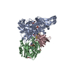
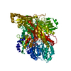
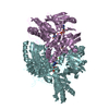
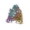
| Title | Crystal structure of the human coronavirus 229E spike protein receptor binding domain in complex with human aminopeptidase N | |||||||||
 Components Components |
| |||||||||
 Keywords Keywords | Hydrolase/Viral protein / coronavirus / spike / receptor / Hydrolase-Viral protein complex | |||||||||
| Function / homology |  Function and homology information Function and homology informationmembrane alanyl aminopeptidase / peptide catabolic process / metalloaminopeptidase activity / endoplasmic reticulum-Golgi intermediate compartment / Metabolism of Angiotensinogen to Angiotensins / aminopeptidase activity / secretory granule membrane / peptide binding / endocytosis involved in viral entry into host cell / metallopeptidase activity ...membrane alanyl aminopeptidase / peptide catabolic process / metalloaminopeptidase activity / endoplasmic reticulum-Golgi intermediate compartment / Metabolism of Angiotensinogen to Angiotensins / aminopeptidase activity / secretory granule membrane / peptide binding / endocytosis involved in viral entry into host cell / metallopeptidase activity / virus receptor activity / signaling receptor activity / angiogenesis / cell differentiation / host cell endoplasmic reticulum-Golgi intermediate compartment membrane / receptor-mediated virion attachment to host cell / external side of plasma membrane / fusion of virus membrane with host plasma membrane / lysosomal membrane / fusion of virus membrane with host endosome membrane / viral envelope / Neutrophil degranulation / virion membrane / proteolysis / extracellular space / zinc ion binding / extracellular exosome / membrane / plasma membrane / cytoplasm Similarity search - Function | |||||||||
| Biological species |  Homo sapiens (human) Homo sapiens (human) Human coronavirus 229E Human coronavirus 229E | |||||||||
| Method |  X-RAY DIFFRACTION / SYNCHROTRON / MOLECULAR REPLACEMENT / Resolution: 3.505 Å X-RAY DIFFRACTION / SYNCHROTRON / MOLECULAR REPLACEMENT / Resolution: 3.505 Å | |||||||||
 Authors Authors | Wong, A.H. / Rini, J.M. | |||||||||
| Funding support |  Canada, 1items Canada, 1items
| |||||||||
 Citation Citation | Journal: Nat Commun / Year: 2017 Title: Receptor-binding loops in alphacoronavirus adaptation and evolution. Authors: Wong, A.H.M. / Tomlinson, A.C.A. / Zhou, D. / Satkunarajah, M. / Chen, K. / Sharon, C. / Desforges, M. / Talbot, P.J. / Rini, J.M. | |||||||||
| History |
|
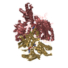
- Structure visualization
| Structure viewer | Molecule: MolmilJmol/JSmol |
|---|
- Downloads & links
-Download
| PDBx/mmCIF format | 6atk.cif.gz | 588.9 KB | Display | PDBx/mmCIF format |
|---|---|---|---|---|
| PDB format | pdb6atk.ent.gz | 475.7 KB | Display | PDB format |
| PDBx/mmJSON format | 6atk.json.gz | Tree view | PDBx/mmJSON format | |
| Others |  Other downloads Other downloads |
-Validation report
| Summary document | 6atk_validation.pdf.gz | 1.6 MB | Display | wwPDB validaton report |
|---|---|---|---|---|
| Full document | 6atk_full_validation.pdf.gz | 1.6 MB | Display | |
| Data in XML | 6atk_validation.xml.gz | 90.9 KB | Display | |
| Data in CIF | 6atk_validation.cif.gz | 122.9 KB | Display | |
| Arichive directory | https://data.pdbj.org/pub/pdb/validation_reports/at/6atkftp://data.pdbj.org/pub/pdb/validation_reports/at/6atk | HTTPS FTP |
-Related structure data
| Related structure data |  4fyqS S: Starting model for refinement |
|---|---|
| Similar structure data |


-Links
 PDBj
PDBj


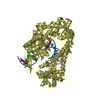
- Assembly
| Deposited unit | 
| ||||||||||||||||||||||||||||||||||||||||||||||||
|---|---|---|---|---|---|---|---|---|---|---|---|---|---|---|---|---|---|---|---|---|---|---|---|---|---|---|---|---|---|---|---|---|---|---|---|---|---|---|---|---|---|---|---|---|---|---|---|---|---|
| 1 | 
| ||||||||||||||||||||||||||||||||||||||||||||||||
| 2 | 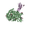
| ||||||||||||||||||||||||||||||||||||||||||||||||
| 3 | 
| ||||||||||||||||||||||||||||||||||||||||||||||||
| Unit cell |
| ||||||||||||||||||||||||||||||||||||||||||||||||
| Noncrystallographic symmetry (NCS) | NCS domain:
NCS domain segments:
|
-Components
| #1: Protein | Mass: 103464.984 Da / Num. of mol.: 3 / Fragment: UNP residues 68-967 Source method: isolated from a genetically manipulated source Source: (gene. exp.) Homo sapiens (human) / Gene: ANPEP, APN, CD13, PEPN / Production host: Homo sapiens (human) / References: UniProt: P15144, membrane alanyl aminopeptidase#2: Protein | Mass: 15868.112 Da / Num. of mol.: 3 / Fragment: UNP residues 294-432 Source method: isolated from a genetically manipulated source Source: (gene. exp.) Human coronavirus 229E / Gene: S, 2 / Production host: Homo sapiens (human) / References: UniProt: P15423#3: Polysaccharide | 2-acetamido-2-deoxy-beta-D-glucopyranose-(1-4)-2-acetamido-2-deoxy-beta-D-glucopyranose Source method: isolated from a genetically manipulated source #4: Sugar | ChemComp-NAG /   Type: D-saccharide, beta linking / Mass: 221.208 Da / Num. of mol.: 15 Type: D-saccharide, beta linking / Mass: 221.208 Da / Num. of mol.: 15Source method: isolated from a genetically manipulated source Formula: C8H15NO6 #5: Chemical |   Mass: 65.409 Da / Num. of mol.: 3 / Source method: obtained synthetically / Formula: Zn Mass: 65.409 Da / Num. of mol.: 3 / Source method: obtained synthetically / Formula: Zn |
|---|
-Experimental details
-Experiment
| Experiment | Method: X-RAY DIFFRACTION / Number of used crystals: 1 |
|---|
- Sample preparation
| Crystal | Density Matthews: 3.12 Å3/Da / Density % sol: 60.61 % |
|---|---|
| Crystal grow | Temperature: 298 K / Method: vapor diffusion, hanging drop Details: 8% PEG 8000, 1mM GSSG, 1mM GSH, 5% Glycerol, 100mM MES, 1ug/mL endo-beta-N-acetylglucosaminidase A |
-Data collection
| Diffraction | Mean temperature: 93 K |
|---|---|
| Diffraction source | Source: SYNCHROTRON / Site: CLSI / Beamline: 08ID-1 / Wavelength: 0.9795 Å |
| Detector | Type: MAR CCD 165 mm / Detector: CCD / Date: Feb 24, 2013 |
| Radiation | Protocol: SINGLE WAVELENGTH / Monochromatic (M) / Laue (L): M / Scattering type: x-ray |
| Radiation wavelength | Wavelength: 0.9795 Å / Relative weight: 1 |
| Reflection | Resolution: 3.5→50 Å / Num. obs: 55989 / % possible obs: 99.6 % / Redundancy: 4.1 % / CC1/2: 0.99 / Rpim(I) all: 0.08 / Net I/σ(I): 10.9 |
| Reflection shell | Resolution: 3.5→3.6 Å / Mean I/σ(I) obs: 2.7 / Num. unique obs: 5490 / CC1/2: 0.68 / Rpim(I) all: 0.33 / % possible all: 99.8 |
- Processing
| Software |
| |||||||||||||||||||||||||||||||||||||||||||||||||||||||||||||||||||||||||||||||||||||||||||||||||||||||||
|---|---|---|---|---|---|---|---|---|---|---|---|---|---|---|---|---|---|---|---|---|---|---|---|---|---|---|---|---|---|---|---|---|---|---|---|---|---|---|---|---|---|---|---|---|---|---|---|---|---|---|---|---|---|---|---|---|---|---|---|---|---|---|---|---|---|---|---|---|---|---|---|---|---|---|---|---|---|---|---|---|---|---|---|---|---|---|---|---|---|---|---|---|---|---|---|---|---|---|---|---|---|---|---|---|---|---|
| Refinement | Method to determine structure: MOLECULAR REPLACEMENT Starting model: 4FYQ Resolution: 3.505→49.795 Å / SU ML: 0.43 / Cross valid method: FREE R-VALUE / σ(F): 1.34 / Phase error: 27.04 / Stereochemistry target values: ML
| |||||||||||||||||||||||||||||||||||||||||||||||||||||||||||||||||||||||||||||||||||||||||||||||||||||||||
| Solvent computation | Shrinkage radii: 0.9 Å / VDW probe radii: 1.11 Å / Solvent model: FLAT BULK SOLVENT MODEL | |||||||||||||||||||||||||||||||||||||||||||||||||||||||||||||||||||||||||||||||||||||||||||||||||||||||||
| Displacement parameters | Biso max: 187.77 Å2 / Biso mean: 96.7655 Å2 / Biso min: 47 Å2 | |||||||||||||||||||||||||||||||||||||||||||||||||||||||||||||||||||||||||||||||||||||||||||||||||||||||||
| Refinement step | Cycle: final / Resolution: 3.505→49.795 Å
| |||||||||||||||||||||||||||||||||||||||||||||||||||||||||||||||||||||||||||||||||||||||||||||||||||||||||
| Refine LS restraints |
| |||||||||||||||||||||||||||||||||||||||||||||||||||||||||||||||||||||||||||||||||||||||||||||||||||||||||
| Refine LS restraints NCS |
| |||||||||||||||||||||||||||||||||||||||||||||||||||||||||||||||||||||||||||||||||||||||||||||||||||||||||
| LS refinement shell | Refine-ID: X-RAY DIFFRACTION / Rfactor Rfree error: 0 / Total num. of bins used: 14
|
