 Movie
Movie Controller


+ Open data

- Basic information
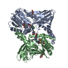
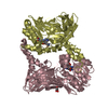
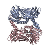
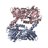
| Entry | Database: PDB / ID: 6arv | ||||||
|---|---|---|---|---|---|---|---|
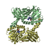
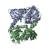
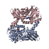
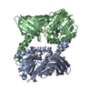
| Title | Crystal structure of CARM1 with Compound 2 and SAH | ||||||
 Components Components | Histone-arginine methyltransferase CARM1 | ||||||
 Keywords Keywords |  TRANSFERASE / protein-inhibitor complex / protein arginine methyltransferase TRANSFERASE / protein-inhibitor complex / protein arginine methyltransferase | ||||||
| Function / homology |  Function and homology information Function and homology information: / histone H3R17 methyltransferase activity / histone H3R2 methyltransferase activity / negative regulation of dendrite development / type I protein arginine methyltransferase / protein-arginine omega-N asymmetric methyltransferase activity / protein methyltransferase activity / histone arginine N-methyltransferase activity / regulation of intracellular estrogen receptor signaling pathway / replication fork reversal ...: / histone H3R17 methyltransferase activity / histone H3R2 methyltransferase activity / negative regulation of dendrite development / type I protein arginine methyltransferase / protein-arginine omega-N asymmetric methyltransferase activity / protein methyltransferase activity / histone arginine N-methyltransferase activity / regulation of intracellular estrogen receptor signaling pathway / replication fork reversal / protein-arginine N-methyltransferase activity / positive regulation of epithelial cell apoptotic process / histone methyltransferase activity / positive regulation of transcription by RNA polymerase I / nuclear replication fork / positive regulation of fat cell differentiation / response to cAMP / RORA activates gene expression / Regulation of lipid metabolism by PPARalpha / TP53 Regulates Transcription of Genes Involved in G2 Cell Cycle Arrest / BMAL1:CLOCK,NPAS2 activates circadian gene expression / Activation of gene expression by SREBF (SREBP) / nuclear receptor coactivator activity / lysine-acetylated histone binding / Heme signaling / Transcriptional activation of mitochondrial biogenesis / PPARA activates gene expression / Cytoprotection by HMOX1 / beta-catenin binding / RMTs methylate histone arginines / Transcriptional regulation of white adipocyte differentiation / Circadian Clock / DNA-binding transcription factor binding / Estrogen-dependent gene expression / transcription coactivator activity / transcription cis-regulatory region binding / positive regulation of cell population proliferation / regulation of DNA-templated transcription / nucleoplasm / nucleus / cytosol / cytoplasmSimilarity search - Function | ||||||
| Biological species |  Homo sapiens (human) Homo sapiens (human) | ||||||
| Method | X-RAY DIFFRACTION / SYNCHROTRON / MOLECULAR REPLACEMENT / Resolution: 2 Å | ||||||
 Authors Authors | Boriack-Sjodin, P.A. / Jin, L. | ||||||
 Citation Citation | Journal: Sci Rep / Year: 2017 Title: Identification of a CARM1 Inhibitor with Potent In Vitro and In Vivo Activity in Preclinical Models of Multiple Myeloma. Authors: Drew, A.E. / Moradei, O. / Jacques, S.L. / Rioux, N. / Boriack-Sjodin, A.P. / Allain, C. / Scott, M.P. / Jin, L. / Raimondi, A. / Handler, J.L. / Ott, H.M. / Kruger, R.G. / McCabe, M.T. / ...Authors: Drew, A.E. / Moradei, O. / Jacques, S.L. / Rioux, N. / Boriack-Sjodin, A.P. / Allain, C. / Scott, M.P. / Jin, L. / Raimondi, A. / Handler, J.L. / Ott, H.M. / Kruger, R.G. / McCabe, M.T. / Sneeringer, C. / Riera, T. / Shapiro, G. / Waters, N.J. / Mitchell, L.H. / Duncan, K.W. / Moyer, M.P. / Copeland, R.A. / Smith, J. / Chesworth, R. / Ribich, S.A. | ||||||
| History |
|
- Structure visualization
| Structure viewer | Molecule: MolmilJmol/JSmol |
|---|
- Downloads & links
-Download
| PDBx/mmCIF format | 6arv.cif.gz | 306.1 KB | Display | PDBx/mmCIF format |
|---|---|---|---|---|
| PDB format | pdb6arv.ent.gz | 246.3 KB | Display | PDB format |
| PDBx/mmJSON format | 6arv.json.gz | Tree view | PDBx/mmJSON format | |
| Others |  Other downloads Other downloads |
-Validation report
| Arichive directory | https://data.pdbj.org/pub/pdb/validation_reports/ar/6arvftp://data.pdbj.org/pub/pdb/validation_reports/ar/6arv | HTTPS FTP |
|---|
-Related structure data

-Links
 PDBj
PDBj


- Assembly
| Deposited unit | 
| ||||||||
|---|---|---|---|---|---|---|---|---|---|
| 1 | 
| ||||||||
| 2 | 
| ||||||||
| Unit cell |
|
-Components
| #1: Protein | Mass: 39623.121 Da / Num. of mol.: 4 / Fragment: Catalytic domain (UNP residues 134-479) Source method: isolated from a genetically manipulated source Source: (gene. exp.) Homo sapiens (human) / Gene: CARM1, PRMT4 / Plasmid: pFastbac / Cell line (production host): Sf9Production host: Insect cell expression vector pTIE1 (others) References: UniProt: Q86X55, type I protein arginine methyltransferase #2: Chemical | ChemComp-SAH / S-Adenosyl-L-homocysteine  Type: L-peptide linking / Mass: 384.411 Da / Num. of mol.: 4 / Source method: obtained synthetically / Formula: C14H20N6O5S Type: L-peptide linking / Mass: 384.411 Da / Num. of mol.: 4 / Source method: obtained synthetically / Formula: C14H20N6O5S#3: Chemical | ChemComp-BW7 / (   Mass: 411.497 Da / Num. of mol.: 4 / Source method: obtained synthetically / Formula: C22H29N5O3 Mass: 411.497 Da / Num. of mol.: 4 / Source method: obtained synthetically / Formula: C22H29N5O3#4: Chemical | ChemComp-GOL / Glycerol  Mass: 92.094 Da / Num. of mol.: 11 / Source method: obtained synthetically / Formula: C3H8O3 Mass: 92.094 Da / Num. of mol.: 11 / Source method: obtained synthetically / Formula: C3H8O3#5: Water | ChemComp-HOH / | Water Mass: 18.015 Da / Num. of mol.: 723 / Source method: isolated from a natural source / Formula: H2O Mass: 18.015 Da / Num. of mol.: 723 / Source method: isolated from a natural source / Formula: H2O |
|---|
-Experimental details
-Experiment
| Experiment | Method: X-RAY DIFFRACTION / Number of used crystals: 1 |
|---|
- Sample preparation
| Crystal | Density Matthews: 2.48 Å3/Da / Density % sol: 50.47 % / Mosaicity: 0 ° |
|---|---|
| Crystal grow | Temperature: 293 K / Method: vapor diffusion, hanging drop / pH: 8.5 Details: 0.2 M Ammonium Sulfate, 0.1 M Tris pH 8.5, 18% w/v PEG3350 |
-Data collection
| Diffraction | Mean temperature: 100 K | |||||||||||||||||||||||||||||||||||||||||||||||||||||||||||||||||||||||||||||||||||||||||||||||||||||||||||||||||||||||||||||||||||||||||||||||||||||||||||||||||||||||||||||||||||||||||||||
|---|---|---|---|---|---|---|---|---|---|---|---|---|---|---|---|---|---|---|---|---|---|---|---|---|---|---|---|---|---|---|---|---|---|---|---|---|---|---|---|---|---|---|---|---|---|---|---|---|---|---|---|---|---|---|---|---|---|---|---|---|---|---|---|---|---|---|---|---|---|---|---|---|---|---|---|---|---|---|---|---|---|---|---|---|---|---|---|---|---|---|---|---|---|---|---|---|---|---|---|---|---|---|---|---|---|---|---|---|---|---|---|---|---|---|---|---|---|---|---|---|---|---|---|---|---|---|---|---|---|---|---|---|---|---|---|---|---|---|---|---|---|---|---|---|---|---|---|---|---|---|---|---|---|---|---|---|---|---|---|---|---|---|---|---|---|---|---|---|---|---|---|---|---|---|---|---|---|---|---|---|---|---|---|---|---|---|---|---|---|---|
| Diffraction source | Source: SYNCHROTRON / Site: APS  / Beamline: 21-ID-G / Wavelength: 0.97856 Å / Beamline: 21-ID-G / Wavelength: 0.97856 Å | |||||||||||||||||||||||||||||||||||||||||||||||||||||||||||||||||||||||||||||||||||||||||||||||||||||||||||||||||||||||||||||||||||||||||||||||||||||||||||||||||||||||||||||||||||||||||||||
| Detector | Type: RAYONIX MX-300 / Detector: CCD / Date: Apr 14, 2012 | |||||||||||||||||||||||||||||||||||||||||||||||||||||||||||||||||||||||||||||||||||||||||||||||||||||||||||||||||||||||||||||||||||||||||||||||||||||||||||||||||||||||||||||||||||||||||||||
| Radiation | Protocol: SINGLE WAVELENGTH / Monochromatic (M) / Laue (L): M / Scattering type: x-ray | |||||||||||||||||||||||||||||||||||||||||||||||||||||||||||||||||||||||||||||||||||||||||||||||||||||||||||||||||||||||||||||||||||||||||||||||||||||||||||||||||||||||||||||||||||||||||||||
| Radiation wavelength | Wavelength: 0.97856 Å / Relative weight: 1 | |||||||||||||||||||||||||||||||||||||||||||||||||||||||||||||||||||||||||||||||||||||||||||||||||||||||||||||||||||||||||||||||||||||||||||||||||||||||||||||||||||||||||||||||||||||||||||||
| Reflection | Resolution: 2→70.78 Å / Num. obs: 105501 / % possible obs: 99.5 % / Redundancy: 4.7 % / Rpim(I) all: 0.045 / Rrim(I) all: 0.097 / Rsym value: 0.086 / Net I/av σ(I): 6.8 / Net I/σ(I): 11.9 | |||||||||||||||||||||||||||||||||||||||||||||||||||||||||||||||||||||||||||||||||||||||||||||||||||||||||||||||||||||||||||||||||||||||||||||||||||||||||||||||||||||||||||||||||||||||||||||
| Reflection shell | Diffraction-ID: 1
|
- Processing
| Software |
| |||||||||||||||||||||||||||||||||||||||||||||
|---|---|---|---|---|---|---|---|---|---|---|---|---|---|---|---|---|---|---|---|---|---|---|---|---|---|---|---|---|---|---|---|---|---|---|---|---|---|---|---|---|---|---|---|---|---|---|
| Refinement | Method to determine structure: MOLECULAR REPLACEMENT / Resolution: 2→70.78 Å / Cor.coef. Fo:Fc: 0.961 / Cor.coef. Fo:Fc free: 0.941 / SU B: 4.461 / SU ML: 0.122 / Cross valid method: THROUGHOUT / σ(F): 0 / ESU R: 0.188 / ESU R Free: 0.165 Details: HYDROGENS HAVE BEEN USED IF PRESENT IN THE INPUT U VALUES : REFINED INDIVIDUALLY
| |||||||||||||||||||||||||||||||||||||||||||||
| Solvent computation | Ion probe radii: 0.8 Å / Shrinkage radii: 0.8 Å / VDW probe radii: 1.2 Å | |||||||||||||||||||||||||||||||||||||||||||||
| Displacement parameters | Biso max: 99.18 Å2 / Biso mean: 30.185 Å2 / Biso min: 14.65 Å2
| |||||||||||||||||||||||||||||||||||||||||||||
| Refinement step | Cycle: final / Resolution: 2→70.78 Å
| |||||||||||||||||||||||||||||||||||||||||||||
| Refine LS restraints |
| |||||||||||||||||||||||||||||||||||||||||||||
| LS refinement shell | Resolution: 2→2.052 Å / Rfactor Rfree error: 0 / Total num. of bins used: 20
|
