 Movie
Movie Controller


+ Open data

- Basic information
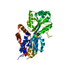
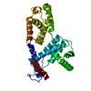
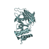
| Entry | Database: PDB / ID: 6amk | ||||||
|---|---|---|---|---|---|---|---|
| Title | Structure of Streptomyces venezuelae BldC-whiI opt complex | ||||||
 Components Components |
| ||||||
 Keywords Keywords | dna binding protein/dna / BldC /  Streptomyces / MerR-like / DNA BINDING PROTEIN / dna binding protein-dna complex Streptomyces / MerR-like / DNA BINDING PROTEIN / dna binding protein-dna complex | ||||||
| Function / homology |  Function and homology information Function and homology information | ||||||
| Biological species |  Streptomyces venezuelae (bacteria) Streptomyces venezuelae (bacteria)synthetic construct (others) | ||||||
| Method | X-RAY DIFFRACTION / SYNCHROTRON / SAD / molecular replacement / Resolution: 3.288 Å | ||||||
 Authors Authors | Schumacher, M.A. | ||||||
 Citation Citation | Journal: Nat Commun / Year: 2018 Title: The MerR-like protein BldC binds DNA direct repeats as cooperative multimers to regulate Streptomyces development. Authors: Schumacher, M.A. / den Hengst, C.D. / Bush, M.J. / Le, T.B.K. / Tran, N.T. / Chandra, G. / Zeng, W. / Travis, B. / Brennan, R.G. / Buttner, M.J. | ||||||
| History |
|
- Structure visualization
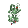
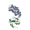
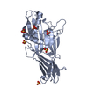
| Structure viewer | Molecule: MolmilJmol/JSmol |
|---|
- Downloads & links
-Download
| PDBx/mmCIF format | 6amk.cif.gz | 99.8 KB | Display | PDBx/mmCIF format |
|---|---|---|---|---|
| PDB format | pdb6amk.ent.gz | 80 KB | Display | PDB format |
| PDBx/mmJSON format | 6amk.json.gz | Tree view | PDBx/mmJSON format | |
| Others |  Other downloads Other downloads |
-Validation report
| Arichive directory | https://data.pdbj.org/pub/pdb/validation_reports/am/6amkftp://data.pdbj.org/pub/pdb/validation_reports/am/6amk | HTTPS FTP |
|---|
-Related structure data


-Links
 PDBj
PDBj


- Assembly
| Deposited unit | 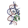
| ||||||||
|---|---|---|---|---|---|---|---|---|---|
| 1 |
| ||||||||
| Unit cell |
|
-Components
| #1: Protein | Mass: 8161.771 Da / Num. of mol.: 2 / Mutation: L43(MSE), L58(MSE) Source method: isolated from a genetically manipulated source Source: (gene. exp.) Streptomyces venezuelae (bacteria) / Gene: AQF52_4259 / Production host: Escherichia coli (E. coli) / References: UniProt: A0A0M7QSG5, UniProt: F2REK9*PLUS#2: DNA chain | | Mass: 6782.389 Da / Num. of mol.: 1 / Source method: obtained synthetically / Source: (synth.) synthetic construct (others) #3: DNA chain | | Mass: 6735.379 Da / Num. of mol.: 1 / Source method: obtained synthetically / Source: (synth.) synthetic construct (others) |
|---|
-Experimental details
-Experiment
| Experiment | Method: X-RAY DIFFRACTION / Number of used crystals: 1 |
|---|
- Sample preparation
| Crystal | Density Matthews: 5.03 Å3/Da / Density % sol: 75.55 % |
|---|---|
| Crystal grow | Temperature: 298 K / Method: vapor diffusion, hanging drop / Details: 30% MPD, 0.1 M sodium acetate pH 5.0, 25% PEG 1500 |
-Data collection
| Diffraction | Mean temperature: 100 K |
|---|---|
| Diffraction source | Source: SYNCHROTRON / Site: ALS  / Beamline: 8.3.1 / Wavelength: 0.979 Å / Beamline: 8.3.1 / Wavelength: 0.979 Å |
| Detector | Type: DECTRIS PILATUS3 S 6M / Detector: PIXEL / Date: Mar 26, 2017 |
| Radiation | Protocol: SINGLE WAVELENGTH / Monochromatic (M) / Laue (L): M / Scattering type: x-ray |
| Radiation wavelength | Wavelength: 0.979 Å / Relative weight: 1 |
| Reflection | Resolution: 3.28→98.9 Å / Num. obs: 16920 / % possible obs: 96.3 % / Observed criterion σ(F): 0 / Redundancy: 9.5 % / Biso Wilson estimate: 114.68 Å2 / CC1/2: 0.99 / Rpim(I) all: 0.055 / Rrim(I) all: 0.14 / Rsym value: 0.116 / Net I/σ(I): 11.7 |
| Reflection shell | Resolution: 3.28→3.46 Å / Redundancy: 9.8 % / CC1/2: 0.665 / Rpim(I) all: 0.67 / Rrim(I) all: 1.59 / Rsym value: 1.43 / % possible all: 97.3 |
-Phasing
| Phasing | Method: molecular replacement |
|---|
- Processing
| Software |
| |||||||||||||||||||||||||||||||||||||||||||||||||||||||||||||||||||||||||||||||||||||||||||
|---|---|---|---|---|---|---|---|---|---|---|---|---|---|---|---|---|---|---|---|---|---|---|---|---|---|---|---|---|---|---|---|---|---|---|---|---|---|---|---|---|---|---|---|---|---|---|---|---|---|---|---|---|---|---|---|---|---|---|---|---|---|---|---|---|---|---|---|---|---|---|---|---|---|---|---|---|---|---|---|---|---|---|---|---|---|---|---|---|---|---|---|---|
| Refinement | Method to determine structure: SAD / Resolution: 3.288→98.9 Å / SU ML: 0.41 / Cross valid method: FREE R-VALUE / σ(F): 0.14 / Phase error: 28.38
| |||||||||||||||||||||||||||||||||||||||||||||||||||||||||||||||||||||||||||||||||||||||||||
| Solvent computation | Shrinkage radii: 0.9 Å / VDW probe radii: 1.11 Å | |||||||||||||||||||||||||||||||||||||||||||||||||||||||||||||||||||||||||||||||||||||||||||
| Displacement parameters | Biso max: 381.62 Å2 / Biso mean: 137.36 Å2 / Biso min: 82.21 Å2 | |||||||||||||||||||||||||||||||||||||||||||||||||||||||||||||||||||||||||||||||||||||||||||
| Refinement step | Cycle: final / Resolution: 3.288→98.9 Å
| |||||||||||||||||||||||||||||||||||||||||||||||||||||||||||||||||||||||||||||||||||||||||||
| Refine LS restraints |
| |||||||||||||||||||||||||||||||||||||||||||||||||||||||||||||||||||||||||||||||||||||||||||
| LS refinement shell | Refine-ID: X-RAY DIFFRACTION / Rfactor Rfree error: 0 / Total num. of bins used: 12
| |||||||||||||||||||||||||||||||||||||||||||||||||||||||||||||||||||||||||||||||||||||||||||
| Refinement TLS params. | Method: refined / Origin x: 4.7044 Å / Origin y: 85.6853 Å / Origin z: 69.6832 Å
| |||||||||||||||||||||||||||||||||||||||||||||||||||||||||||||||||||||||||||||||||||||||||||
| Refinement TLS group |
|
