 Movie
Movie Controller

[English] 日本語
 Yorodumi
- PDB-6akv: Crystal structure of LysB4, the endolysin from Bacillus cereus-ta... -
+ Open data

- Basic information
| Entry | Database: PDB / ID: 6akv | ||||||
|---|---|---|---|---|---|---|---|
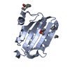
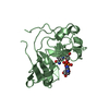
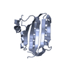
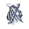
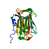
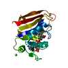
| Title | Crystal structure of LysB4, the endolysin from Bacillus cereus-targeting bacteriophage B4 | ||||||
 Components Components | LysB4 | ||||||
 Keywords Keywords |  HYDROLASE / endolysin / LAS type enzyme / L-Alanoyl D-Glutamate endopeptidase HYDROLASE / endolysin / LAS type enzyme / L-Alanoyl D-Glutamate endopeptidase | ||||||
| Function / homology |  Function and homology information Function and homology information | ||||||
| Biological species |  Bacillus phage B4 (virus) Bacillus phage B4 (virus) | ||||||
| Method | X-RAY DIFFRACTION / SYNCHROTRON / MOLECULAR REPLACEMENT / Resolution: 2.4 Å | ||||||
 Authors Authors | Hong, S. / Ha, N.-C. | ||||||
 Citation Citation | Journal: Mol. Cells / Year: 2019 Title: Crystal Structure of LysB4, an Endolysin fromBacillus cereus-Targeting Bacteriophage B4. Authors: Hong, S. / Son, B. / Ryu, S. / Ha, N.C. | ||||||
| History |
|
- Structure visualization
| Structure viewer | Molecule: MolmilJmol/JSmol |
|---|
- Downloads & links
-Download
| PDBx/mmCIF format | 6akv.cif.gz | 47.9 KB | Display | PDBx/mmCIF format |
|---|---|---|---|---|
| PDB format | pdb6akv.ent.gz | 29.9 KB | Display | PDB format |
| PDBx/mmJSON format | 6akv.json.gz | Tree view | PDBx/mmJSON format | |
| Others |  Other downloads Other downloads |
-Validation report
| Arichive directory | https://data.pdbj.org/pub/pdb/validation_reports/ak/6akvftp://data.pdbj.org/pub/pdb/validation_reports/ak/6akv | HTTPS FTP |
|---|
-Related structure data
| Related structure data |  2vo9S S: Starting model for refinement |
|---|---|
| Similar structure data |


-Links
 PDBj
PDBj
- Assembly
| Deposited unit | 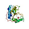
| ||||||||
|---|---|---|---|---|---|---|---|---|---|
| 1 |
| ||||||||
| Unit cell |
|
-Components
| #1: Protein | Mass: 30083.279 Da / Num. of mol.: 1 Source method: isolated from a genetically manipulated source Source: (gene. exp.) Bacillus phage B4 (virus) / Gene: lysB4, BCB4_0006 / Production host:  Escherichia coli BL21(DE3) (bacteria) / Strain (production host): BL21 (DE3) / References: UniProt: H9NAL3 Escherichia coli BL21(DE3) (bacteria) / Strain (production host): BL21 (DE3) / References: UniProt: H9NAL3 |
|---|---|
| #2: Chemical | ChemComp-ZN /   Mass: 65.409 Da / Num. of mol.: 1 / Source method: obtained synthetically / Formula: Zn Mass: 65.409 Da / Num. of mol.: 1 / Source method: obtained synthetically / Formula: Zn |
| #3: Chemical | ChemComp-SO4 / Sulfate  Mass: 96.063 Da / Num. of mol.: 1 / Source method: obtained synthetically / Formula: SO4 Mass: 96.063 Da / Num. of mol.: 1 / Source method: obtained synthetically / Formula: SO4 |
| #4: Chemical | ChemComp-CL / Chloride  Mass: 35.453 Da / Num. of mol.: 1 / Source method: obtained synthetically / Formula: Cl Mass: 35.453 Da / Num. of mol.: 1 / Source method: obtained synthetically / Formula: Cl |
| #5: Water | ChemComp-HOH / Water Mass: 18.015 Da / Num. of mol.: 23 / Source method: isolated from a natural source / Formula: H2O Mass: 18.015 Da / Num. of mol.: 23 / Source method: isolated from a natural source / Formula: H2O |
-Experimental details
-Experiment
| Experiment | Method: X-RAY DIFFRACTION / Number of used crystals: 1 |
|---|
- Sample preparation
| Crystal | Density Matthews: 2.04 Å3/Da / Density % sol: 39.83 % |
|---|---|
| Crystal grow | Temperature: 287.15 K / Method: vapor diffusion, hanging drop / pH: 6.5 Details: 2.0 M ammonium sulfate, 0.1 M Bis-Tris pH 6.5, 2% polyethylene glycol monomethyl ether 550 (PEGMME 550), 8 mM Tris(2-carboxyethyl)phosphine (TCEP) |
-Data collection
| Diffraction | Mean temperature: 100 K |
|---|---|
| Diffraction source | Source: SYNCHROTRON / Site: PAL/PLS  / Beamline: 7A (6B, 6C1) / Wavelength: 0.97934 Å / Beamline: 7A (6B, 6C1) / Wavelength: 0.97934 Å |
| Detector | Type: ADSC QUANTUM 270 / Detector: CCD / Date: May 29, 2017 |
| Radiation | Protocol: SINGLE WAVELENGTH / Monochromatic (M) / Laue (L): M / Scattering type: x-ray |
| Radiation wavelength | Wavelength: 0.97934 Å / Relative weight: 1 |
| Reflection | Resolution: 2.4→50 Å / Num. obs: 8863 / % possible obs: 91.5 % / Redundancy: 4.9 % / Rmerge(I) obs: 0.057 / Rpim(I) all: 0.025 / Rrim(I) all: 0.063 / Net I/σ(I): 19.66 |
| Reflection shell | Resolution: 2.4→2.44 Å / Redundancy: 2.9 % / Rmerge(I) obs: 0.351 / Mean I/σ(I) obs: 2.36 / Num. unique obs: 449 / CC1/2: 0.353 / Rpim(I) all: 0.222 / Rrim(I) all: 0.419 / % possible all: 92.4 |
- Processing
| Software |
| ||||||||||||||||||||||||||||||||||||||||||
|---|---|---|---|---|---|---|---|---|---|---|---|---|---|---|---|---|---|---|---|---|---|---|---|---|---|---|---|---|---|---|---|---|---|---|---|---|---|---|---|---|---|---|---|
| Refinement | Method to determine structure: MOLECULAR REPLACEMENT Starting model: 2VO9 Resolution: 2.4→28.092 Å / SU ML: 0.29 / Cross valid method: FREE R-VALUE / σ(F): 1.51 / Phase error: 22.2
| ||||||||||||||||||||||||||||||||||||||||||
| Solvent computation | Shrinkage radii: 0.9 Å / VDW probe radii: 1.11 Å | ||||||||||||||||||||||||||||||||||||||||||
| Refinement step | Cycle: LAST / Resolution: 2.4→28.092 Å
| ||||||||||||||||||||||||||||||||||||||||||
| Refine LS restraints |
| ||||||||||||||||||||||||||||||||||||||||||
| LS refinement shell |
|
