 Movie
Movie Controller

[English] 日本語
 Yorodumi
- PDB-6ahi: Crystal structure of O-acetylserine dependent cystathionine beta-... -
+ Open data

- Basic information
| Entry | Database: PDB / ID: 6ahi | |||||||||
|---|---|---|---|---|---|---|---|---|---|---|
| Title | Crystal structure of O-acetylserine dependent cystathionine beta-synthase from Helicobacter pylori. | |||||||||
 Components Components | Cysteine synthase | |||||||||
 Keywords Keywords | TRANSFERASE | |||||||||
| Function / homology |  Function and homology informationcystathionine beta-synthase activity / cysteine synthase / cysteine synthase activity / cysteine biosynthetic process / cysteine biosynthetic process from serine / cytoplasm Function and homology informationcystathionine beta-synthase activity / cysteine synthase / cysteine synthase activity / cysteine biosynthetic process / cysteine biosynthetic process from serine / cytoplasmSimilarity search - Function | |||||||||
| Biological species |   Helicobacter pylori (bacteria) Helicobacter pylori (bacteria) | |||||||||
| Method | X-RAY DIFFRACTION / SYNCHROTRON / MOLECULAR REPLACEMENT / Resolution: 1.9 Å | |||||||||
 Authors Authors | Tarique, F.K. / Devi, S. / Rehman, S.A.A. | |||||||||
 Citation Citation | Journal: Mol.Microbiol. / Year: 2019 Title: Identification and characterization of Helicobacter pylori O-acetylserine-dependent cystathionine beta-synthase, a distinct member of the PLP-II family. Authors: Devi, S. / Tarique, K.F. / Ali, M.F. / Abdul Rehman, S.A. / Gourinath, S. | |||||||||
| History |
|
- Structure visualization
| Structure viewer | Molecule: MolmilJmol/JSmol |
|---|
- Downloads & links
-Download
| PDBx/mmCIF format | 6ahi.cif.gz | 249.7 KB | Display | PDBx/mmCIF format |
|---|---|---|---|---|
| PDB format | pdb6ahi.ent.gz | 200 KB | Display | PDB format |
| PDBx/mmJSON format | 6ahi.json.gz | Tree view | PDBx/mmJSON format | |
| Others |  Other downloads Other downloads |
-Validation report
| Arichive directory | https://data.pdbj.org/pub/pdb/validation_reports/ah/6ahiftp://data.pdbj.org/pub/pdb/validation_reports/ah/6ahi | HTTPS FTP |
|---|
-Related structure data
| Related structure data | 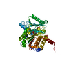 1z7wS S: Starting model for refinement |
|---|---|
| Similar structure data |


-Links
 PDBj
PDBj
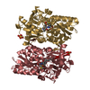
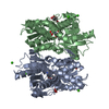
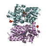
- Assembly
| Deposited unit | 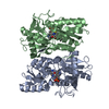
| ||||||||||||||||||
|---|---|---|---|---|---|---|---|---|---|---|---|---|---|---|---|---|---|---|---|
| 1 |
| ||||||||||||||||||
| Unit cell |
| ||||||||||||||||||
| Noncrystallographic symmetry (NCS) | NCS domain:
NCS domain segments: Component-ID: 0 / Ens-ID: 1 / Beg auth comp-ID: MET / Beg label comp-ID: MET / End auth comp-ID: LEU / End label comp-ID: LEU / Refine code: 0 / Auth seq-ID: 1 - 306 / Label seq-ID: 1 - 306
|
-Components
| #1: Protein | / CSase / O-acetylserine (thiol)-lyase / OAS-TL / O-acetylserine sulfhydrylase Mass: 34273.926 Da / Num. of mol.: 2 Source method: isolated from a genetically manipulated source Source: (gene. exp.) Helicobacter pylori (strain ATCC 700392 / 26695) (bacteria)Strain: ATCC 700392 / 26695 / Gene: cysM, cysK, HP_0107 Production host: Escherichia coli 'BL21-Gold(DE3)pLysS AG' (bacteria)Strain (production host): BL21(DE3) / References: UniProt: P56067, cysteine synthase#2: Chemical | ChemComp-MET / | Methionine  Type: L-peptide linking / Mass: 149.211 Da / Num. of mol.: 1 / Source method: obtained synthetically / Formula: C5H11NO2S Type: L-peptide linking / Mass: 149.211 Da / Num. of mol.: 1 / Source method: obtained synthetically / Formula: C5H11NO2S#3: Water | ChemComp-HOH / | Water Mass: 18.015 Da / Num. of mol.: 132 / Source method: isolated from a natural source / Formula: H2O Mass: 18.015 Da / Num. of mol.: 132 / Source method: isolated from a natural source / Formula: H2O |
|---|
-Experimental details
-Experiment
| Experiment | Method: X-RAY DIFFRACTION / Number of used crystals: 1 |
|---|
- Sample preparation
| Crystal | Density Matthews: 2.14 Å3/Da / Density % sol: 42.46 % |
|---|---|
| Crystal grow | Temperature: 289 K / Method: vapor diffusion, hanging drop Details: Peg20K-Peg550MME 15%, 0.09M Amino acids mixture, 0.1M Mes-Imadazole buffer pH 6.5 |
-Data collection
| Diffraction | Mean temperature: 100 K |
|---|---|
| Diffraction source | Source: SYNCHROTRON / Site: ESRF  / Beamline: BM14 / Wavelength: 0.978 Å / Beamline: BM14 / Wavelength: 0.978 Å |
| Detector | Type: MAR scanner 300 mm plate / Detector: IMAGE PLATE / Date: Dec 3, 2011 |
| Radiation | Protocol: SINGLE WAVELENGTH / Monochromatic (M) / Laue (L): M / Scattering type: x-ray |
| Radiation wavelength | Wavelength: 0.978 Å / Relative weight: 1 |
| Reflection | Resolution: 1.9→50 Å / Num. obs: 45368 / % possible obs: 100 % / Redundancy: 16.3 % / Rmerge(I) obs: 0.117 / Net I/σ(I): 47 |
| Reflection shell | Resolution: 1.9→1.93 Å / Rmerge(I) obs: 0.862 / Num. unique obs: 2244 |
- Processing
| Software |
| ||||||||||||||||||||||||||||||||||||||||||||||||||||||||||||||||||||||||||||||||||||||||||||||||||||||||||||||||||||||||||||||||||||||||||||||||||||||||||||||||||||||||||||||||||||||
|---|---|---|---|---|---|---|---|---|---|---|---|---|---|---|---|---|---|---|---|---|---|---|---|---|---|---|---|---|---|---|---|---|---|---|---|---|---|---|---|---|---|---|---|---|---|---|---|---|---|---|---|---|---|---|---|---|---|---|---|---|---|---|---|---|---|---|---|---|---|---|---|---|---|---|---|---|---|---|---|---|---|---|---|---|---|---|---|---|---|---|---|---|---|---|---|---|---|---|---|---|---|---|---|---|---|---|---|---|---|---|---|---|---|---|---|---|---|---|---|---|---|---|---|---|---|---|---|---|---|---|---|---|---|---|---|---|---|---|---|---|---|---|---|---|---|---|---|---|---|---|---|---|---|---|---|---|---|---|---|---|---|---|---|---|---|---|---|---|---|---|---|---|---|---|---|---|---|---|---|---|---|---|---|
| Refinement | Method to determine structure: MOLECULAR REPLACEMENT Starting model: 1Z7W Resolution: 1.9→48.12 Å / Cor.coef. Fo:Fc: 0.962 / Cor.coef. Fo:Fc free: 0.955 / SU B: 8.444 / SU ML: 0.11 / Cross valid method: THROUGHOUT / ESU R: 0.16 / ESU R Free: 0.135 / Details: HYDROGENS HAVE BEEN ADDED IN THE RIDING POSITIONS
| ||||||||||||||||||||||||||||||||||||||||||||||||||||||||||||||||||||||||||||||||||||||||||||||||||||||||||||||||||||||||||||||||||||||||||||||||||||||||||||||||||||||||||||||||||||||
| Solvent computation | Ion probe radii: 0.7 Å / Shrinkage radii: 0.7 Å / VDW probe radii: 1 Å | ||||||||||||||||||||||||||||||||||||||||||||||||||||||||||||||||||||||||||||||||||||||||||||||||||||||||||||||||||||||||||||||||||||||||||||||||||||||||||||||||||||||||||||||||||||||
| Displacement parameters | Biso mean: 37.455 Å2
| ||||||||||||||||||||||||||||||||||||||||||||||||||||||||||||||||||||||||||||||||||||||||||||||||||||||||||||||||||||||||||||||||||||||||||||||||||||||||||||||||||||||||||||||||||||||
| Refinement step | Cycle: 1 / Resolution: 1.9→48.12 Å
| ||||||||||||||||||||||||||||||||||||||||||||||||||||||||||||||||||||||||||||||||||||||||||||||||||||||||||||||||||||||||||||||||||||||||||||||||||||||||||||||||||||||||||||||||||||||
| Refine LS restraints |
|