| Entry | Database: PDB / ID: 6aam
|
|---|
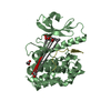
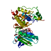
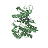
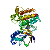
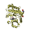
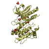
| Title | Crystal structure of TYK2 in complex with peficitinib |
|---|
 Components Components | Non-receptor tyrosine-protein kinase TYK2 |
|---|
 Keywords Keywords | TRANSFERASE/INHIBITOR /  protein kinase / TRANSFERASE-INHIBITOR complex protein kinase / TRANSFERASE-INHIBITOR complex |
|---|
| Function / homology |  Function and homology information Function and homology information
type III interferon-mediated signaling pathway / interleukin-12 receptor complex / interleukin-23 receptor complex / Interleukin-23 signaling / positive regulation of T-helper 17 type immune response / type 1 angiotensin receptor binding / positive regulation of NK T cell proliferation / interleukin-12-mediated signaling pathway / Interleukin-12 signaling / Interleukin-35 Signalling ...type III interferon-mediated signaling pathway / interleukin-12 receptor complex / interleukin-23 receptor complex / Interleukin-23 signaling / positive regulation of T-helper 17 type immune response / type 1 angiotensin receptor binding / positive regulation of NK T cell proliferation / interleukin-12-mediated signaling pathway / Interleukin-12 signaling / Interleukin-35 Signalling / Interleukin-27 signaling / IL-6-type cytokine receptor ligand interactions / growth hormone receptor binding / positive regulation of natural killer cell proliferation / Other interleukin signaling / extrinsic component of plasma membrane / Interleukin-20 family signaling / type I interferon-mediated signaling pathway / Interleukin-6 signaling / positive regulation of interleukin-17 production / MAPK3 (ERK1) activation / Interleukin-10 signaling / MAPK1 (ERK2) activation / cell surface receptor signaling pathway via JAK-STAT / Regulation of IFNA/IFNB signaling / growth hormone receptor signaling pathway via JAK-STAT / type II interferon-mediated signaling pathway / positive regulation of T cell proliferation / Signaling by CSF3 (G-CSF) / Evasion by RSV of host interferon responses / non-specific protein-tyrosine kinase / positive regulation of receptor signaling pathway via JAK-STAT / non-membrane spanning protein tyrosine kinase activity / Signaling by ALK fusions and activated point mutants / Inactivation of CSF3 (G-CSF) signaling / cytoplasmic side of plasma membrane / cellular response to virus / cytokine-mediated signaling pathway / positive regulation of protein localization to nucleus / positive regulation of type II interferon production / Interferon alpha/beta signaling / protein tyrosine kinase activity / Interleukin-4 and Interleukin-13 signaling / Potential therapeutics for SARS / cell differentiation / cytoskeleton / intracellular signal transduction / immune response / protein phosphorylation / SARS-CoV-2 activates/modulates innate and adaptive immune responses / extracellular exosome / ATP binding / nucleus / plasma membrane / cytosol / cytoplasmSimilarity search - Function Tyrosine-protein kinase, non-receptor, TYK2, N-terminal / Tyrosine-protein kinase, non-receptor Jak/Tyk2 / JAK, FERM F2 lobe domain / FERM F1 lobe ubiquitin-like domain / JAK1-3/TYK2, pleckstrin homology-like domain / Jak1 pleckstrin homology-like domain / FERM F2 acyl-CoA binding protein-like domain / FERM F1 ubiquitin-like domain / FERM superfamily, second domain / FERM domain ...Tyrosine-protein kinase, non-receptor, TYK2, N-terminal / Tyrosine-protein kinase, non-receptor Jak/Tyk2 / JAK, FERM F2 lobe domain / FERM F1 lobe ubiquitin-like domain / JAK1-3/TYK2, pleckstrin homology-like domain / Jak1 pleckstrin homology-like domain / FERM F2 acyl-CoA binding protein-like domain / FERM F1 ubiquitin-like domain / FERM superfamily, second domain / FERM domain / FERM domain profile. / Band 4.1 domain / Band 4.1 homologues / Src homology 2 domains / SH2 domain / SH2 domain superfamily / Tyrosine-protein kinase, catalytic domain / Tyrosine kinase, catalytic domain / Tyrosine protein kinases specific active-site signature. / Tyrosine-protein kinase, active site / Protein tyrosine and serine/threonine kinase / Serine-threonine/tyrosine-protein kinase, catalytic domain / Transferase(Phosphotransferase) domain 1 / Transferase(Phosphotransferase); domain 1 / Phosphorylase Kinase; domain 1 / Phosphorylase Kinase; domain 1 / Protein kinase, ATP binding site / Protein kinases ATP-binding region signature. / Protein kinase domain profile. / Protein kinase domain / Protein kinase-like domain superfamily / 2-Layer Sandwich / Orthogonal Bundle / Mainly Alpha / Alpha BetaSimilarity search - Domain/homology |
|---|
| Biological species |  Homo sapiens (human) Homo sapiens (human) |
|---|
| Method | X-RAY DIFFRACTION / SYNCHROTRON / MOLECULAR REPLACEMENT / Resolution: 1.98 Å |
|---|
 Authors Authors | Nomura, N. / Tomimoto, Y. |
|---|
 Citation Citation | Journal: Bioorg. Med. Chem. / Year: 2018
Title: Discovery and structural characterization of peficitinib (ASP015K) as a novel and potent JAK inhibitor
Authors: Hamaguchi, H. / Amano, Y. / Moritomo, A. / Shirakami, S. / Nakajima, Y. / Nakai, K. / Nomura, N. / Ito, M. / Higashi, Y. / Inoue, T. |
|---|
| History | | Deposition | Jul 18, 2018 | Deposition site: PDBJ / Processing site: PDBJ |
|---|
| Revision 1.0 | Aug 15, 2018 | Provider: repository / Type: Initial release |
|---|
| Revision 1.1 | Sep 12, 2018 | Group: Data collection / Database references / Category: citation
Item: _citation.journal_abbrev / _citation.pdbx_database_id_PubMed |
|---|
| Revision 1.2 | Oct 24, 2018 | Group: Data collection / Database references / Category: citation
Item: _citation.journal_volume / _citation.page_first / _citation.page_last |
|---|
| Revision 1.3 | Mar 27, 2024 | Group: Data collection / Database references / Category: chem_comp_atom / chem_comp_bond / database_2
Item: _database_2.pdbx_DOI / _database_2.pdbx_database_accession |
|---|
|
|---|
 Movie
Movie Controller


 Open data
 Basic information
 Structure visualization
 Downloads & links
 Other downloads
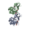


 PDBj
PDBj


 Assembly
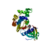


 Mass: 326.393 Da / Num. of mol.: 1 / Source method: obtained synthetically / Formula: C18H22N4O2
 Mass: 18.015 Da / Num. of mol.: 45 / Source method: isolated from a natural source / Formula: H2O
 Sample preparation
 / Beamline: AR-NE3A / Wavelength: 1 Å
 Processing