 Movie
Movie Controller


+ Open data

- Basic information
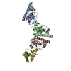
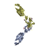
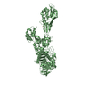
| Entry | Database: PDB / ID: 6a9y | ||||||
|---|---|---|---|---|---|---|---|
| Title | The crystal structure of Mu homology domain of SGIP1 | ||||||
 Components Components | SH3-containing GRB2-like protein 3-interacting protein 1 | ||||||
 Keywords Keywords |  ENDOCYTOSIS / SGIP1 / dimer / disulfide bond ENDOCYTOSIS / SGIP1 / dimer / disulfide bond | ||||||
| Function / homology |  Function and homology information Function and homology informationpositive regulation of feeding behavior / AP-2 adaptor complex / clathrin-dependent endocytosis / clathrin-coated vesicle / response to dietary excess / energy homeostasis / clathrin-coated pit / phospholipid binding / SH3 domain binding / positive regulation of receptor-mediated endocytosis ...positive regulation of feeding behavior / AP-2 adaptor complex / clathrin-dependent endocytosis / clathrin-coated vesicle / response to dietary excess / energy homeostasis / clathrin-coated pit / phospholipid binding / SH3 domain binding / positive regulation of receptor-mediated endocytosis / Cargo recognition for clathrin-mediated endocytosis / Clathrin-mediated endocytosis / microtubule binding / plasma membrane / cytosol / cytoplasmSimilarity search - Function | ||||||
| Biological species |  Homo sapiens (human) Homo sapiens (human) | ||||||
| Method | X-RAY DIFFRACTION / SYNCHROTRON / MOLECULAR REPLACEMENT / Resolution: 2.7 Å | ||||||
 Authors Authors | Feng, Y. / Liu, X. | ||||||
 Citation Citation | Journal: Biochem. Biophys. Res. Commun. / Year: 2018 Title: SGIP1 dimerizes via intermolecular disulfide bond in mu HD domain during cellular endocytosis. Authors: Zhang, Y. / Feng, Y. / Xin, Y. / Liu, X. | ||||||
| History |
|
- Structure visualization
| Structure viewer | Molecule: MolmilJmol/JSmol |
|---|
- Downloads & links
-Download
| PDBx/mmCIF format | 6a9y.cif.gz | 71.1 KB | Display | PDBx/mmCIF format |
|---|---|---|---|---|
| PDB format | pdb6a9y.ent.gz | 50.9 KB | Display | PDB format |
| PDBx/mmJSON format | 6a9y.json.gz | Tree view | PDBx/mmJSON format | |
| Others |  Other downloads Other downloads |
-Validation report
| Arichive directory | https://data.pdbj.org/pub/pdb/validation_reports/a9/6a9yftp://data.pdbj.org/pub/pdb/validation_reports/a9/6a9y | HTTPS FTP |
|---|
-Related structure data
| Related structure data | 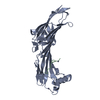 5awuS S: Starting model for refinement |
|---|---|
| Similar structure data |


-Links
 PDBj
PDBj
- Assembly
| Deposited unit | 
| ||||||||
|---|---|---|---|---|---|---|---|---|---|
| 1 | 
| ||||||||
| Unit cell |
| ||||||||
| Components on special symmetry positions |
|

-Components
| #1: Protein | Mass: 31269.523 Da / Num. of mol.: 1 / Fragment: Mu homology domain Source method: isolated from a genetically manipulated source Source: (gene. exp.) Homo sapiens (human) / Gene: SGIP1 / Production host:  Enterobacteria phage L1 (virus) / References: UniProt: Q9BQI5 Enterobacteria phage L1 (virus) / References: UniProt: Q9BQI5 |
|---|---|
| #2: Water | ChemComp-HOH / Water Mass: 18.015 Da / Num. of mol.: 61 / Source method: isolated from a natural source / Formula: H2O Mass: 18.015 Da / Num. of mol.: 61 / Source method: isolated from a natural source / Formula: H2O |
-Experimental details
-Experiment
| Experiment | Method: X-RAY DIFFRACTION / Number of used crystals: 1 |
|---|
- Sample preparation
| Crystal | Density Matthews: 3.01 Å3/Da / Density % sol: 59.2 % |
|---|---|
| Crystal grow | Temperature: 293 K / Method: vapor diffusion / Details: 0.1M Tris, pH 8.0, 3.0 M NaCl |
-Data collection
| Diffraction | Mean temperature: 100 K |
|---|---|
| Diffraction source | Source: SYNCHROTRON / Site: SSRF  / Beamline: BL17U / Wavelength: 0.9792 Å / Beamline: BL17U / Wavelength: 0.9792 Å |
| Detector | Type: MARMOSAIC 225 mm CCD / Detector: CCD / Date: Jul 10, 2017 |
| Radiation | Protocol: SINGLE WAVELENGTH / Monochromatic (M) / Laue (L): M / Scattering type: x-ray |
| Radiation wavelength | Wavelength: 0.9792 Å / Relative weight: 1 |
| Reflection | Resolution: 2.7→30 Å / Num. obs: 10898 / % possible obs: 99.73 % / Redundancy: 6.5 % / Rsym value: 0.09 / Net I/σ(I): 35.3 |
| Reflection shell | Resolution: 2.7→2.97 Å / Num. unique obs: 565 / Rsym value: 0.45 |
- Processing
| Software |
| |||||||||||||||||||||||||||||||||||
|---|---|---|---|---|---|---|---|---|---|---|---|---|---|---|---|---|---|---|---|---|---|---|---|---|---|---|---|---|---|---|---|---|---|---|---|---|
| Refinement | Method to determine structure: MOLECULAR REPLACEMENT Starting model: 5AWU Resolution: 2.7→29.912 Å / SU ML: 0.36 / Cross valid method: FREE R-VALUE / σ(F): 1.36 / Phase error: 31.25
| |||||||||||||||||||||||||||||||||||
| Solvent computation | Shrinkage radii: 0.9 Å / VDW probe radii: 1.11 Å | |||||||||||||||||||||||||||||||||||
| Refinement step | Cycle: LAST / Resolution: 2.7→29.912 Å
| |||||||||||||||||||||||||||||||||||
| Refine LS restraints |
| |||||||||||||||||||||||||||||||||||
| LS refinement shell |
|
