 Movie
Movie Controller

[English] 日本語
 Yorodumi
- PDB-6a9p: Crystal structure of the human glial fibrillary acidic protein 1B... -
+ Open data

- Basic information
| Entry | Database: PDB / ID: 6a9p | ||||||
|---|---|---|---|---|---|---|---|
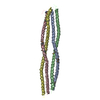
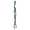
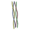
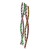
| Title | Crystal structure of the human glial fibrillary acidic protein 1B domain | ||||||
 Components Components | Glial fibrillary acidic protein | ||||||
 Keywords Keywords | STRUCTURAL PROTEIN / Glial fibrillary acidic protein. GFAP / Intermediate filament / IF / Alexander disease / 1B domain | ||||||
| Function / homology |  Function and homology informationregulation of chaperone-mediated autophagy / intermediate filament organization / intermediate filament / intermediate filament cytoskeleton / regulation of protein-containing complex assembly / Nuclear signaling by ERBB4 / structural constituent of cytoskeleton / Chaperone Mediated Autophagy / identical protein binding / cytosol / cytoplasm Function and homology informationregulation of chaperone-mediated autophagy / intermediate filament organization / intermediate filament / intermediate filament cytoskeleton / regulation of protein-containing complex assembly / Nuclear signaling by ERBB4 / structural constituent of cytoskeleton / Chaperone Mediated Autophagy / identical protein binding / cytosol / cytoplasmSimilarity search - Function | ||||||
| Biological species |  Homo sapiens (human) Homo sapiens (human) | ||||||
| Method | X-RAY DIFFRACTION / SYNCHROTRON / MOLECULAR REPLACEMENT / Resolution: 2.51 Å | ||||||
 Authors Authors | Jin, M.S. / Kim, B. / Kim, S. | ||||||
| Funding support |  Korea, Republic Of, 1items Korea, Republic Of, 1items
| ||||||
 Citation Citation | Journal: Biochem.Biophys.Res.Commun. / Year: 2018 Title: Crystal structure of the human glial fibrillary acidic protein 1B domain Authors: Kim, B. / Kim, S. / Jin, M.S. | ||||||
| History |
|
- Structure visualization
| Structure viewer | Molecule: MolmilJmol/JSmol |
|---|
- Downloads & links
-Download
| PDBx/mmCIF format | 6a9p.cif.gz | 350.1 KB | Display | PDBx/mmCIF format |
|---|---|---|---|---|
| PDB format | pdb6a9p.ent.gz | 292.3 KB | Display | PDB format |
| PDBx/mmJSON format | 6a9p.json.gz | Tree view | PDBx/mmJSON format | |
| Others |  Other downloads Other downloads |
-Validation report
| Arichive directory | https://data.pdbj.org/pub/pdb/validation_reports/a9/6a9pftp://data.pdbj.org/pub/pdb/validation_reports/a9/6a9p | HTTPS FTP |
|---|
-Related structure data
| Related structure data |  3uf1S S: Starting model for refinement |
|---|---|
| Similar structure data |


-Links
 PDBj
PDBj


- Assembly
| Deposited unit | 
| ||||||||
|---|---|---|---|---|---|---|---|---|---|
| 1 | 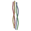
| ||||||||
| 2 | 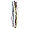
| ||||||||
| Unit cell |
|
-Components
| #1: Protein | / GFAP Mass: 12604.056 Da / Num. of mol.: 8 Source method: isolated from a genetically manipulated source Source: (gene. exp.) Homo sapiens (human) / Gene: GFAP / Production host:  Escherichia coli (E. coli) / References: UniProt: P14136 Escherichia coli (E. coli) / References: UniProt: P14136#2: Water | ChemComp-HOH / | Water Mass: 18.015 Da / Num. of mol.: 132 / Source method: isolated from a natural source / Formula: H2O Mass: 18.015 Da / Num. of mol.: 132 / Source method: isolated from a natural source / Formula: H2O |
|---|
-Experimental details
-Experiment
| Experiment | Method: X-RAY DIFFRACTION / Number of used crystals: 1 |
|---|
- Sample preparation
| Crystal | Density Matthews: 2.78 Å3/Da / Density % sol: 55.7 % |
|---|---|
| Crystal grow | Temperature: 295 K / Method: vapor diffusion, sitting drop / pH: 8 Details: 100 mM Tris-HCl pH 8.0, 400 mM NaCl and 22% (w/v) PEG10,000 |
-Data collection
| Diffraction | Mean temperature: 80 K |
|---|---|
| Diffraction source | Source: SYNCHROTRON / Site: PAL/PLS / Beamline: 7A (6B, 6C1) / Wavelength: 1 Å |
| Detector | Type: ADSC QUANTUM 270 / Detector: CCD / Date: Jun 8, 2016 |
| Radiation | Protocol: SINGLE WAVELENGTH / Monochromatic (M) / Laue (L): M / Scattering type: x-ray |
| Radiation wavelength | Wavelength: 1 Å / Relative weight: 1 |
| Reflection | Resolution: 2.5→50 Å / Num. obs: 29465 / % possible obs: 94.9 % / Redundancy: 1.9 % / Rmerge(I) obs: 0.062 / Net I/σ(I): 18.2 |
| Reflection shell | Resolution: 2.5→2.59 Å / Rmerge(I) obs: 0.614 |
- Processing
| Software |
| ||||||||||||||||||||||||||||||||||||||||||||||||||||||||||||||||||||||||||||||||||||||||||||||||||||||||||||||||||||||||||||||||||||||||||||||||||||||||||||||||||||||||||
|---|---|---|---|---|---|---|---|---|---|---|---|---|---|---|---|---|---|---|---|---|---|---|---|---|---|---|---|---|---|---|---|---|---|---|---|---|---|---|---|---|---|---|---|---|---|---|---|---|---|---|---|---|---|---|---|---|---|---|---|---|---|---|---|---|---|---|---|---|---|---|---|---|---|---|---|---|---|---|---|---|---|---|---|---|---|---|---|---|---|---|---|---|---|---|---|---|---|---|---|---|---|---|---|---|---|---|---|---|---|---|---|---|---|---|---|---|---|---|---|---|---|---|---|---|---|---|---|---|---|---|---|---|---|---|---|---|---|---|---|---|---|---|---|---|---|---|---|---|---|---|---|---|---|---|---|---|---|---|---|---|---|---|---|---|---|---|---|---|---|---|---|
| Refinement | Method to determine structure: MOLECULAR REPLACEMENT Starting model: 3UF1 Resolution: 2.51→50 Å / Cor.coef. Fo:Fc: 0.886 / Cor.coef. Fo:Fc free: 0.853 / SU B: 31.932 / SU ML: 0.331 / Cross valid method: THROUGHOUT / ESU R Free: 0.407 / Details: HYDROGENS HAVE BEEN ADDED IN THE RIDING POSITIONS
| ||||||||||||||||||||||||||||||||||||||||||||||||||||||||||||||||||||||||||||||||||||||||||||||||||||||||||||||||||||||||||||||||||||||||||||||||||||||||||||||||||||||||||
| Solvent computation | Ion probe radii: 0.8 Å / Shrinkage radii: 0.8 Å / VDW probe radii: 1.2 Å | ||||||||||||||||||||||||||||||||||||||||||||||||||||||||||||||||||||||||||||||||||||||||||||||||||||||||||||||||||||||||||||||||||||||||||||||||||||||||||||||||||||||||||
| Displacement parameters | Biso mean: 57.085 Å2
| ||||||||||||||||||||||||||||||||||||||||||||||||||||||||||||||||||||||||||||||||||||||||||||||||||||||||||||||||||||||||||||||||||||||||||||||||||||||||||||||||||||||||||
| Refinement step | Cycle: 1 / Resolution: 2.51→50 Å
| ||||||||||||||||||||||||||||||||||||||||||||||||||||||||||||||||||||||||||||||||||||||||||||||||||||||||||||||||||||||||||||||||||||||||||||||||||||||||||||||||||||||||||
| Refine LS restraints |
| ||||||||||||||||||||||||||||||||||||||||||||||||||||||||||||||||||||||||||||||||||||||||||||||||||||||||||||||||||||||||||||||||||||||||||||||||||||||||||||||||||||||||||
| LS refinement shell | Resolution: 2.508→2.573 Å / Total num. of bins used: 20
|
