 Movie
Movie Controller

[English] 日本語
 Yorodumi
- PDB-6a4s: Crystal structure of peptidase E with ordered active site loop fr... -
+ Open data

- Basic information
| Entry | Database: PDB / ID: 6a4s | ||||||
|---|---|---|---|---|---|---|---|
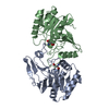
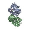
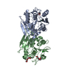
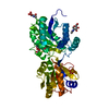
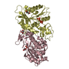
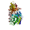
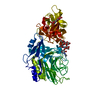
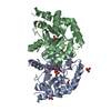
| Title | Crystal structure of peptidase E with ordered active site loop from Salmonella enterica | ||||||
 Components Components | Peptidase E Dipeptidase E Dipeptidase E | ||||||
 Keywords Keywords | HYDROLASE / S51 peptidase / peptidase E / dimer / active site loop | ||||||
| Function / homology |  Function and homology informationdipeptidase E / dipeptidase activity / serine-type peptidase activity / proteolysis / cytoplasm Function and homology informationdipeptidase E / dipeptidase activity / serine-type peptidase activity / proteolysis / cytoplasmSimilarity search - Function | ||||||
| Biological species |  Salmonella typhimurium (bacteria) Salmonella typhimurium (bacteria) | ||||||
| Method | X-RAY DIFFRACTION / SYNCHROTRON / MOLECULAR REPLACEMENT / Resolution: 1.9 Å | ||||||
 Authors Authors | Yadav, P. / Chandravanshi, K. / Goyal, V.D. / Singh, R. / Kumar, A. / Gokhale, S.M. / Makde, R.D. | ||||||
 Citation Citation | Journal: FEBS Lett. / Year: 2018 Title: Structure of Asp-bound peptidase E from Salmonella enterica: Active site at dimer interface illuminates Asp recognition. Authors: Yadav, P. / Goyal, V.D. / Gaur, N.K. / Kumar, A. / Gokhale, S.M. / Makde, R.D. | ||||||
| History |
|
- Structure visualization
| Structure viewer | Molecule: MolmilJmol/JSmol |
|---|
- Downloads & links
-Download
| PDBx/mmCIF format | 6a4s.cif.gz | 198.5 KB | Display | PDBx/mmCIF format |
|---|---|---|---|---|
| PDB format | pdb6a4s.ent.gz | 156.4 KB | Display | PDB format |
| PDBx/mmJSON format | 6a4s.json.gz | Tree view | PDBx/mmJSON format | |
| Others |  Other downloads Other downloads |
-Validation report
| Arichive directory | https://data.pdbj.org/pub/pdb/validation_reports/a4/6a4sftp://data.pdbj.org/pub/pdb/validation_reports/a4/6a4s | HTTPS FTP |
|---|
-Related structure data
| Related structure data |  6a4rC 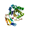 1fy2S S: Starting model for refinement C: citing same article ( |
|---|---|
| Similar structure data |


-Links
 PDBj
PDBj

- Assembly
| Deposited unit | 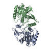
| ||||||||
|---|---|---|---|---|---|---|---|---|---|
| 1 |
| ||||||||
| Unit cell |
|
-Components
| #1: Protein | Dipeptidase E / Alpha-aspartyl dipeptidase / Asp-specific dipeptidase / Dipeptidase E Mass: 28596.221 Da / Num. of mol.: 2 Source method: isolated from a genetically manipulated source Source: (gene. exp.) Salmonella typhimurium (strain LT2 / SGSC1412 / ATCC 700720) (bacteria)Strain: LT2 / SGSC1412 / ATCC 700720 / Gene: pepE, STM4190 / Production host: Escherichia coli BL21(DE3) (bacteria) / Strain (production host): BL21(DE3) / References: UniProt: P36936, dipeptidase E#2: Water | ChemComp-HOH / | Water Mass: 18.015 Da / Num. of mol.: 307 / Source method: isolated from a natural source / Formula: H2O Mass: 18.015 Da / Num. of mol.: 307 / Source method: isolated from a natural source / Formula: H2O |
|---|
-Experimental details
-Experiment
| Experiment | Method: X-RAY DIFFRACTION / Number of used crystals: 1 |
|---|
- Sample preparation
| Crystal | Density Matthews: 2.17 Å3/Da / Density % sol: 43.19 % |
|---|---|
| Crystal grow | Temperature: 294 K / Method: microbatch / pH: 6.5 Details: 0.1 M Bis-tris pH6.5, 0.2 M Sodium Chloride, 25 % PEG 3350 PH range: 5-6.5 |
-Data collection
| Diffraction | Mean temperature: 100 K |
|---|---|
| Diffraction source | Source: SYNCHROTRON / Site: RRCAT INDUS-2  / Beamline: PX-BL21 / Wavelength: 0.97949 Å / Beamline: PX-BL21 / Wavelength: 0.97949 Å |
| Detector | Type: MAR scanner 345 mm plate / Detector: IMAGE PLATE / Date: Jun 6, 2018 / Details: mirrors |
| Radiation | Monochromator: Si111 / Protocol: SINGLE WAVELENGTH / Monochromatic (M) / Laue (L): M / Scattering type: x-ray |
| Radiation wavelength | Wavelength: 0.97949 Å / Relative weight: 1 |
| Reflection | Resolution: 1.9→45.77 Å / Num. obs: 38789 / % possible obs: 99.4 % / Redundancy: 3.2 % / Biso Wilson estimate: 20.7 Å2 / CC1/2: 0.998 / Rmerge(I) obs: 0.053 / Rpim(I) all: 0.035 / Rrim(I) all: 0.064 / Net I/σ(I): 16.9 |
| Reflection shell | Resolution: 1.9→1.94 Å / Redundancy: 3.1 % / Rmerge(I) obs: 0.388 / Mean I/σ(I) obs: 3.2 / Num. unique obs: 2495 / CC1/2: 0.886 / Rpim(I) all: 0.254 / Rrim(I) all: 0.465 / % possible all: 99.3 |
- Processing
| Software |
| ||||||||||||||||||||||||||||||||||||||||||||||||||||||||||||||||||||||||||||||||||||||||||||||||||
|---|---|---|---|---|---|---|---|---|---|---|---|---|---|---|---|---|---|---|---|---|---|---|---|---|---|---|---|---|---|---|---|---|---|---|---|---|---|---|---|---|---|---|---|---|---|---|---|---|---|---|---|---|---|---|---|---|---|---|---|---|---|---|---|---|---|---|---|---|---|---|---|---|---|---|---|---|---|---|---|---|---|---|---|---|---|---|---|---|---|---|---|---|---|---|---|---|---|---|---|
| Refinement | Method to determine structure: MOLECULAR REPLACEMENT Starting model: 1FY2 Resolution: 1.9→41.461 Å / SU ML: 0.17 / Cross valid method: FREE R-VALUE / σ(F): 1.34 / Phase error: 21.26
| ||||||||||||||||||||||||||||||||||||||||||||||||||||||||||||||||||||||||||||||||||||||||||||||||||
| Solvent computation | Shrinkage radii: 0.9 Å / VDW probe radii: 1.11 Å | ||||||||||||||||||||||||||||||||||||||||||||||||||||||||||||||||||||||||||||||||||||||||||||||||||
| Displacement parameters | Biso mean: 23 Å2 | ||||||||||||||||||||||||||||||||||||||||||||||||||||||||||||||||||||||||||||||||||||||||||||||||||
| Refinement step | Cycle: LAST / Resolution: 1.9→41.461 Å
| ||||||||||||||||||||||||||||||||||||||||||||||||||||||||||||||||||||||||||||||||||||||||||||||||||
| Refine LS restraints |
| ||||||||||||||||||||||||||||||||||||||||||||||||||||||||||||||||||||||||||||||||||||||||||||||||||
| LS refinement shell |
| ||||||||||||||||||||||||||||||||||||||||||||||||||||||||||||||||||||||||||||||||||||||||||||||||||
| Refinement TLS params. | Method: refined / Origin x: -12.3172 Å / Origin y: -0.4181 Å / Origin z: -21.272 Å
| ||||||||||||||||||||||||||||||||||||||||||||||||||||||||||||||||||||||||||||||||||||||||||||||||||
| Refinement TLS group | Selection details: all |
