 Movie
Movie Controller

[English] 日本語
 Yorodumi
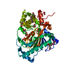
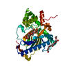
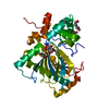
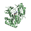
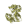
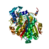
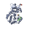
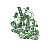
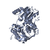
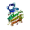
- PDB-6a2e: Apo structure of the Kdo hydroxylase KdoO, a non-heme Fe(II) alph... -
+ Open data

- Basic information
| Entry | Database: PDB / ID: 6a2e | ||||||
|---|---|---|---|---|---|---|---|
| Title | Apo structure of the Kdo hydroxylase KdoO, a non-heme Fe(II) alphaketoglutarate dependent dioxygenase | ||||||
 Components Components | Kdo hydroxylase, KdoO | ||||||
 Keywords Keywords |  BIOSYNTHETIC PROTEIN / Fe(II)/O2/alphaketoglutarate dependent dioxygenase / KDO2-lipid A dioxygenase / deoxysugar dioxygenase / LPS biosynthesis BIOSYNTHETIC PROTEIN / Fe(II)/O2/alphaketoglutarate dependent dioxygenase / KDO2-lipid A dioxygenase / deoxysugar dioxygenase / LPS biosynthesis | ||||||
| Function / homology | Kdo hydroxylase / 3-deoxy-D-manno-oct-2-ulosonic acid (Kdo) hydroxylase / ACETATE ION / 3-deoxy-D-manno-oct-2-ulosonic acid (Kdo) hydroxylase Function and homology information Function and homology information | ||||||
| Biological species |  Methylacidiphilum infernorum (bacteria) Methylacidiphilum infernorum (bacteria) | ||||||
| Method | X-RAY DIFFRACTION / SYNCHROTRON / SAD / Resolution: 1.939 Å | ||||||
 Authors Authors | Chung, H.S. / Pemble, C.W. / Joo, S.H. | ||||||
| Funding support |  Korea, Republic Of, 1items Korea, Republic Of, 1items
| ||||||
 Citation Citation | Journal: J. Mol. Biol. / Year: 2018 Title: Biochemical and Structural Insights into an Fe(II)/ alpha-Ketoglutarate/O2-Dependent Dioxygenase, Kdo 3-Hydroxylase (KdoO). Authors: Joo, S.H. / Pemble, C.W. / Yang, E.G. / Raetz, C.R.H. / Chung, H.S. | ||||||
| History |
|
- Structure visualization
| Structure viewer | Molecule: MolmilJmol/JSmol |
|---|
- Downloads & links
-Download
| PDBx/mmCIF format | 6a2e.cif.gz | 137.7 KB | Display | PDBx/mmCIF format |
|---|---|---|---|---|
| PDB format | pdb6a2e.ent.gz | 105.9 KB | Display | PDB format |
| PDBx/mmJSON format | 6a2e.json.gz | Tree view | PDBx/mmJSON format | |
| Others |  Other downloads Other downloads |
-Validation report
| Arichive directory | https://data.pdbj.org/pub/pdb/validation_reports/a2/6a2eftp://data.pdbj.org/pub/pdb/validation_reports/a2/6a2e | HTTPS FTP |
|---|
-Related structure data


-Links
 PDBj
PDBj- Assembly
| Deposited unit | 
| ||||||||
|---|---|---|---|---|---|---|---|---|---|
| 1 |
| ||||||||
| Unit cell |
|
-Components
| #1: Protein | Mass: 36239.316 Da / Num. of mol.: 1 Source method: isolated from a genetically manipulated source Source: (gene. exp.) Methylacidiphilum infernorum (isolate V4) (bacteria)Strain: V4 / Gene: Minf_1012 / Production host: Escherichia coli (E. coli) / References: UniProt: B3DUR4 | ||||||
|---|---|---|---|---|---|---|---|
| #2: Chemical | Acetate  Mass: 59.044 Da / Num. of mol.: 3 / Source method: obtained synthetically / Formula: C2H3O2 Mass: 59.044 Da / Num. of mol.: 3 / Source method: obtained synthetically / Formula: C2H3O2#3: Chemical | ChemComp-SO4 / | Sulfate  Mass: 96.063 Da / Num. of mol.: 1 / Source method: obtained synthetically / Formula: SO4 Mass: 96.063 Da / Num. of mol.: 1 / Source method: obtained synthetically / Formula: SO4#4: Chemical | ChemComp-PG4 / | Polyethylene glycol  Mass: 194.226 Da / Num. of mol.: 1 / Source method: obtained synthetically / Formula: C8H18O5 / Comment: precipitant*YM Mass: 194.226 Da / Num. of mol.: 1 / Source method: obtained synthetically / Formula: C8H18O5 / Comment: precipitant*YM#5: Water | ChemComp-HOH / | Water Mass: 18.015 Da / Num. of mol.: 252 / Source method: isolated from a natural source / Formula: H2O Mass: 18.015 Da / Num. of mol.: 252 / Source method: isolated from a natural source / Formula: H2O |
-Experimental details
-Experiment
| Experiment | Method: X-RAY DIFFRACTION / Number of used crystals: 1 |
|---|
- Sample preparation
| Crystal | Density Matthews: 2.19 Å3/Da / Density % sol: 43.91 % |
|---|---|
| Crystal grow | Temperature: 288 K / Method: vapor diffusion, sitting drop / pH: 4.5 Details: 0.1M Sodium acetate, 200mM Lithium sulfate, 50% v/v PEG 400 |
-Data collection
| Diffraction | Mean temperature: 100 K | |||||||||||||||||||||||||||||||||||||||||||||||||||||||||||||||||||||||||||||||||||||||||||||||||||||||||||||||||||||||||||||||||||||||||||||||||||
|---|---|---|---|---|---|---|---|---|---|---|---|---|---|---|---|---|---|---|---|---|---|---|---|---|---|---|---|---|---|---|---|---|---|---|---|---|---|---|---|---|---|---|---|---|---|---|---|---|---|---|---|---|---|---|---|---|---|---|---|---|---|---|---|---|---|---|---|---|---|---|---|---|---|---|---|---|---|---|---|---|---|---|---|---|---|---|---|---|---|---|---|---|---|---|---|---|---|---|---|---|---|---|---|---|---|---|---|---|---|---|---|---|---|---|---|---|---|---|---|---|---|---|---|---|---|---|---|---|---|---|---|---|---|---|---|---|---|---|---|---|---|---|---|---|---|---|---|---|
| Diffraction source | Source: SYNCHROTRON / Site: APS  / Beamline: 22-BM / Wavelength: 1 Å / Beamline: 22-BM / Wavelength: 1 Å | |||||||||||||||||||||||||||||||||||||||||||||||||||||||||||||||||||||||||||||||||||||||||||||||||||||||||||||||||||||||||||||||||||||||||||||||||||
| Detector | Type: MARMOSAIC 225 mm CCD / Detector: CCD / Date: Jun 23, 2011 | |||||||||||||||||||||||||||||||||||||||||||||||||||||||||||||||||||||||||||||||||||||||||||||||||||||||||||||||||||||||||||||||||||||||||||||||||||
| Radiation | Monochromator: Si(111) / Protocol: SINGLE WAVELENGTH / Monochromatic (M) / Laue (L): M / Scattering type: x-ray | |||||||||||||||||||||||||||||||||||||||||||||||||||||||||||||||||||||||||||||||||||||||||||||||||||||||||||||||||||||||||||||||||||||||||||||||||||
| Radiation wavelength | Wavelength: 1 Å / Relative weight: 1 | |||||||||||||||||||||||||||||||||||||||||||||||||||||||||||||||||||||||||||||||||||||||||||||||||||||||||||||||||||||||||||||||||||||||||||||||||||
| Reflection | Resolution: 1.939→50 Å / Num. obs: 24341 / % possible obs: 99.3 % / Redundancy: 7 % / Biso Wilson estimate: 18.21 Å2 / Rmerge(I) obs: 0.097 / Χ2: 1.735 / Net I/σ(I): 8.5 | |||||||||||||||||||||||||||||||||||||||||||||||||||||||||||||||||||||||||||||||||||||||||||||||||||||||||||||||||||||||||||||||||||||||||||||||||||
| Reflection shell | Diffraction-ID: 1
|
- Processing
| Software |
| |||||||||||||||||||||||||||||||||||||||||||||||||||||||||||||||||||||||||||
|---|---|---|---|---|---|---|---|---|---|---|---|---|---|---|---|---|---|---|---|---|---|---|---|---|---|---|---|---|---|---|---|---|---|---|---|---|---|---|---|---|---|---|---|---|---|---|---|---|---|---|---|---|---|---|---|---|---|---|---|---|---|---|---|---|---|---|---|---|---|---|---|---|---|---|---|---|
| Refinement | Method to determine structure: SAD / Resolution: 1.939→41.605 Å / SU ML: 0.14 / Cross valid method: FREE R-VALUE / σ(F): 0 / Phase error: 19.19
| |||||||||||||||||||||||||||||||||||||||||||||||||||||||||||||||||||||||||||
| Solvent computation | Shrinkage radii: 0.9 Å / VDW probe radii: 1.11 Å | |||||||||||||||||||||||||||||||||||||||||||||||||||||||||||||||||||||||||||
| Displacement parameters | Biso max: 80.08 Å2 / Biso mean: 23.8371 Å2 / Biso min: 8.35 Å2 | |||||||||||||||||||||||||||||||||||||||||||||||||||||||||||||||||||||||||||
| Refinement step | Cycle: final / Resolution: 1.939→41.605 Å
| |||||||||||||||||||||||||||||||||||||||||||||||||||||||||||||||||||||||||||
| Refine LS restraints |
| |||||||||||||||||||||||||||||||||||||||||||||||||||||||||||||||||||||||||||
| LS refinement shell | Refine-ID: X-RAY DIFFRACTION / Rfactor Rfree error: 0 / Total num. of bins used: 9
| |||||||||||||||||||||||||||||||||||||||||||||||||||||||||||||||||||||||||||
| Refinement TLS params. | Method: refined / Refine-ID: X-RAY DIFFRACTION
| |||||||||||||||||||||||||||||||||||||||||||||||||||||||||||||||||||||||||||
| Refinement TLS group |
|
