 Movie
Movie Controller


+ Open data

- Basic information
| Entry | Database: PDB / ID: 5zrt | ||||||
|---|---|---|---|---|---|---|---|
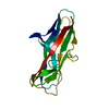
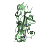
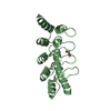
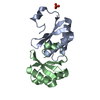
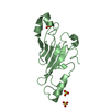
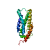
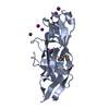
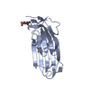
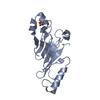
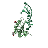
| Title | Crystal structure of human C1ORF123 protein | ||||||
 Components Components | UPF0587 protein C1orf123 | ||||||
 Keywords Keywords | UNKNOWN FUNCTION /  human hypothetical protein human hypothetical protein | ||||||
| Function / homology | CXXC motif containing zinc binding protein, eukaryotic / CXXC motif containing zinc binding protein, eukaryotic / zinc ion binding / BETA-MERCAPTOETHANOL / CXXC motif containing zinc binding protein Function and homology information Function and homology information | ||||||
| Biological species |  Homo sapiens (human) Homo sapiens (human) | ||||||
| Method | X-RAY DIFFRACTION / SYNCHROTRON / MOLECULAR REPLACEMENT / Resolution: 1.9 Å | ||||||
 Authors Authors | Rahaman, S.N.A. / Yusop, J.M. / Mohamed-Hussein, Z.A. / Wan Mohd, A. / Ho, K.L. / Teh, A.H. / Waterman, J. / Ng, C.L. | ||||||
| Funding support |  Malaysia, 1items Malaysia, 1items
| ||||||
 Citation Citation | Journal: Peerj / Year: 2018 Title: Crystal structure and functional analysis of human C1ORF123. Authors: A Rahaman, S.N. / Mat Yusop, J. / Mohamed-Hussein, Z.A. / Aizat, W.M. / Ho, K.L. / Teh, A.H. / Waterman, J. / Tan, B.K. / Tan, H.L. / Li, A.Y. / Chen, E.S. / Ng, C.L. #1: Journal: Acta Crystallogr F Struct Biol Commun / Year: 2016 Title: Cloning, expression, purification, crystallization and X-ray crystallographic analysis of recombinant human C1ORF123 protein Authors: Rhaman, S.N. / Yusop, M.J. / Mohamed-Hussein, Z.A. / Ho, K.L. / Teh, A.H. / Waterman, J. / Ng, C.L. | ||||||
| History |
|
- Structure visualization
| Structure viewer | Molecule: MolmilJmol/JSmol |
|---|
- Downloads & links
-Download
| PDBx/mmCIF format | 5zrt.cif.gz | 149.8 KB | Display | PDBx/mmCIF format |
|---|---|---|---|---|
| PDB format | pdb5zrt.ent.gz | 116.4 KB | Display | PDB format |
| PDBx/mmJSON format | 5zrt.json.gz | Tree view | PDBx/mmJSON format | |
| Others |  Other downloads Other downloads |
-Validation report
| Arichive directory | https://data.pdbj.org/pub/pdb/validation_reports/zr/5zrtftp://data.pdbj.org/pub/pdb/validation_reports/zr/5zrt | HTTPS FTP |
|---|
-Related structure data
| Related structure data |  1zsoS S: Starting model for refinement |
|---|---|
| Similar structure data |
-Links
 PDBj
PDBj


- Assembly
| Deposited unit | 
| ||||||||||||||||||
|---|---|---|---|---|---|---|---|---|---|---|---|---|---|---|---|---|---|---|---|
| 1 | 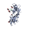
| ||||||||||||||||||
| 2 | 
| ||||||||||||||||||
| Unit cell |
| ||||||||||||||||||
| Noncrystallographic symmetry (NCS) | NCS domain:
NCS domain segments: Component-ID: 0 / Ens-ID: 1 / Beg auth comp-ID: MET / Beg label comp-ID: MET / End auth comp-ID: CYS / End label comp-ID: CYS / Refine code: 0 / Auth seq-ID: 1 - 160 / Label seq-ID: 21 - 180
|
-Components
-Protein , 1 types, 2 molecules AB
| #1: Protein | Mass: 20239.797 Da / Num. of mol.: 2 Source method: isolated from a genetically manipulated source Source: (gene. exp.) Homo sapiens (human) / Gene: C1orf123 / Production host:  Escherichia coli (E. coli) / References: UniProt: Q9NWV4 Escherichia coli (E. coli) / References: UniProt: Q9NWV4 |
|---|
-Non-polymers , 5 types, 190 molecules 


| #2: Chemical | ChemComp-GOL / Glycerol Mass: 92.094 Da / Num. of mol.: 6 / Source method: obtained synthetically / Formula: C3H8O3 Mass: 92.094 Da / Num. of mol.: 6 / Source method: obtained synthetically / Formula: C3H8O3#3: Chemical | Chloride Mass: 35.453 Da / Num. of mol.: 2 / Source method: obtained synthetically / Formula: Cl Mass: 35.453 Da / Num. of mol.: 2 / Source method: obtained synthetically / Formula: Cl#4: Chemical |  Mass: 65.409 Da / Num. of mol.: 2 / Source method: obtained synthetically / Formula: Zn Mass: 65.409 Da / Num. of mol.: 2 / Source method: obtained synthetically / Formula: Zn#5: Chemical | ChemComp-BME / | 2-Mercaptoethanol Mass: 78.133 Da / Num. of mol.: 1 / Source method: obtained synthetically / Formula: C2H6OS Mass: 78.133 Da / Num. of mol.: 1 / Source method: obtained synthetically / Formula: C2H6OS#6: Water | ChemComp-HOH / | WaterMass: 18.015 Da / Num. of mol.: 179 / Source method: isolated from a natural source / Formula: H2O |
|---|
-Experimental details
-Experiment
| Experiment | Method: X-RAY DIFFRACTION / Number of used crystals: 1 |
|---|
- Sample preparation
| Crystal | Density Matthews: 2.28 Å3/Da / Density % sol: 45.95 % / Description: Long rod |
|---|---|
| Crystal grow | Temperature: 293 K / Method: vapor diffusion / pH: 6.5 Details: 0.2M magnesium chloride hexahydrate, 0.1M sodium citrate tribasic buffer pH 6.5, 20% polyethylene glycol 3350 PH range: 6.5 |
-Data collection
| Diffraction | Mean temperature: 100 K |
|---|---|
| Diffraction source | Source: SYNCHROTRON / Site: Diamond  / Beamline: I02 / Wavelength: 0.9797 Å / Beamline: I02 / Wavelength: 0.9797 Å |
| Detector | Type: DECTRIS PILATUS3 S 6M / Detector: PIXEL / Date: Oct 10, 2014 |
| Radiation | Protocol: SINGLE WAVELENGTH / Monochromatic (M) / Laue (L): M / Scattering type: x-ray |
| Radiation wavelength | Wavelength: 0.9797 Å / Relative weight: 1 |
| Reflection | Resolution: 1.9→30.9 Å / Num. obs: 29180 / % possible obs: 98.1 % / Observed criterion σ(I): 3 / Redundancy: 5.5 % / Rmerge(I) obs: 0.06 / Rsym value: 0.065 / Net I/σ(I): 13.8 |
| Reflection shell | Resolution: 1.9→1.94 Å / Redundancy: 5.3 % / Mean I/σ(I) obs: 3 / Num. unique obs: 10058 / Rsym value: 0.526 / % possible all: 97.2 |
- Processing
| Software |
| |||||||||||||||||||||||||||||||||||||||||||||||||||||||||||||||||||||||||||
|---|---|---|---|---|---|---|---|---|---|---|---|---|---|---|---|---|---|---|---|---|---|---|---|---|---|---|---|---|---|---|---|---|---|---|---|---|---|---|---|---|---|---|---|---|---|---|---|---|---|---|---|---|---|---|---|---|---|---|---|---|---|---|---|---|---|---|---|---|---|---|---|---|---|---|---|---|
| Refinement | Method to determine structure: MOLECULAR REPLACEMENT Starting model: 1ZSO Resolution: 1.9→30.9 Å / Cor.coef. Fo:Fc: 0.966 / Cor.coef. Fo:Fc free: 0.944 / SU B: 6.102 / SU ML: 0.095 / Cross valid method: THROUGHOUT / σ(F): 0 / ESU R: 0.14 / ESU R Free: 0.136 / Stereochemistry target values: MAXIMUM LIKELIHOOD / Details: U VALUES : WITH TLS ADDED
| |||||||||||||||||||||||||||||||||||||||||||||||||||||||||||||||||||||||||||
| Solvent computation | Ion probe radii: 0.8 Å / Shrinkage radii: 0.8 Å / VDW probe radii: 1.2 Å / Solvent model: MASK | |||||||||||||||||||||||||||||||||||||||||||||||||||||||||||||||||||||||||||
| Displacement parameters | Biso max: 125.73 Å2 / Biso mean: 46.861 Å2 / Biso min: 20.52 Å2
| |||||||||||||||||||||||||||||||||||||||||||||||||||||||||||||||||||||||||||
| Refinement step | Cycle: final / Resolution: 1.9→30.9 Å
| |||||||||||||||||||||||||||||||||||||||||||||||||||||||||||||||||||||||||||
| Refine LS restraints |
| |||||||||||||||||||||||||||||||||||||||||||||||||||||||||||||||||||||||||||
| Refine LS restraints NCS | Ens-ID: 1 / Number: 185 / Refine-ID: X-RAY DIFFRACTION / Type: interatomic distance / Rms dev position: 0.2 Å / Weight position: 0.05
| |||||||||||||||||||||||||||||||||||||||||||||||||||||||||||||||||||||||||||
| LS refinement shell | Resolution: 1.9→1.949 Å / Rfactor Rfree error: 0 / Total num. of bins used: 20
| |||||||||||||||||||||||||||||||||||||||||||||||||||||||||||||||||||||||||||
| Refinement TLS params. | Method: refined / Refine-ID: X-RAY DIFFRACTION
| |||||||||||||||||||||||||||||||||||||||||||||||||||||||||||||||||||||||||||
| Refinement TLS group |
|
