 Movie
Movie Controller

[English] 日本語
 Yorodumi
- PDB-5zka: Crystal structure of N-acetylneuraminate lyase from Fusobacterium... -
+ Open data

- Basic information
| Entry | Database: PDB / ID: 5zka | |||||||||||||||
|---|---|---|---|---|---|---|---|---|---|---|---|---|---|---|---|---|
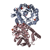
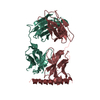
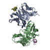
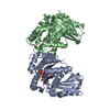
| Title | Crystal structure of N-acetylneuraminate lyase from Fusobacterium nucleatum complexed with Pyruvate | |||||||||||||||
 Components Components | (N-acetylneuraminate lyase ) x 2 ) x 2 | |||||||||||||||
 Keywords Keywords | LYASE / N-acetylneuraminate lyase / Sialic acid catabolism / TIM-barrel / Schiff base | |||||||||||||||
| Function / homology |  Function and homology informationN-acetylneuraminate lyase / N-acetylneuraminate lyase activity / N-acetylneuraminate catabolic process / carbohydrate metabolic process / cytosol Function and homology informationN-acetylneuraminate lyase / N-acetylneuraminate lyase activity / N-acetylneuraminate catabolic process / carbohydrate metabolic process / cytosolSimilarity search - Function | |||||||||||||||
| Biological species |  Fusobacterium nucleatum subsp. nucleatum ATCC 25586 (bacteria) Fusobacterium nucleatum subsp. nucleatum ATCC 25586 (bacteria) | |||||||||||||||
| Method | X-RAY DIFFRACTION / SYNCHROTRON / MOLECULAR REPLACEMENT / Resolution: 1.76 Å | |||||||||||||||
 Authors Authors | Kumar, J.P. / Rao, H. / Nayak, V. / Ramaswamy, S. | |||||||||||||||
| Funding support |  India, 4items India, 4items
| |||||||||||||||
 Citation Citation | Journal: Acta Crystallogr F Struct Biol Commun / Year: 2018 Title: Crystal structures and kinetics of N-acetylneuraminate lyase from Fusobacterium nucleatum Authors: Kumar, J.P. / Rao, H. / Nayak, V. / Ramaswamy, S. #1: Journal: Biochemistry / Year: 2013Title: Structural basis for substrate specificity and mechanism of N-acetyl-D-neuraminic acid lyase from Pasteurella multocida. Authors: Huynh, N. / Aye, A. / Li, Y. / Yu, H. / Cao, H. / Tiwari, V.K. / Shin, D.W. / Chen, X. / Fisher, A.J. | |||||||||||||||
| History |
|
- Structure visualization
| Structure viewer | Molecule: MolmilJmol/JSmol |
|---|
- Downloads & links
-Download
| PDBx/mmCIF format | 5zka.cif.gz | 245.8 KB | Display | PDBx/mmCIF format |
|---|---|---|---|---|
| PDB format | pdb5zka.ent.gz | 196.4 KB | Display | PDB format |
| PDBx/mmJSON format | 5zka.json.gz | Tree view | PDBx/mmJSON format | |
| Others |  Other downloads Other downloads |
-Validation report
| Arichive directory | https://data.pdbj.org/pub/pdb/validation_reports/zk/5zkaftp://data.pdbj.org/pub/pdb/validation_reports/zk/5zka | HTTPS FTP |
|---|
-Related structure data
| Related structure data |  5zjmC  4imcS S: Starting model for refinement C: citing same article ( |
|---|---|
| Similar structure data |


-Links
 PDBj
PDBj- Assembly
| Deposited unit | 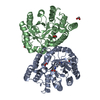
| ||||||||
|---|---|---|---|---|---|---|---|---|---|
| 1 |
| ||||||||
| Unit cell |
|
-Components
| #1: Protein | / Neu5Ac lyase / N-acetylneuraminate pyruvate-lyase / N-acetylneuraminic acid aldolase / Sialate ...Neu5Ac lyase / N-acetylneuraminate pyruvate-lyase / N-acetylneuraminic acid aldolase / Sialate lyase / Sialic acid aldolase / Sialic acid lyase Mass: 34983.371 Da / Num. of mol.: 1 Source method: isolated from a genetically manipulated source Details: Pyruvate covalently bound through a Schiff base to Lys161 Source: (gene. exp.) Fusobacterium nucleatum subsp. nucleatum ATCC 25586 (bacteria)Strain: ATCC 25586 / Gene: nanA / Plasmid: pET300/NT-DEST / Details (production host): From Invitrogen / Production host: Escherichia coli BL21(DE3) (bacteria) / Strain (production host): BL21(DE3) / References: UniProt: Q8RDN6, N-acetylneuraminate lyase | ||||||
|---|---|---|---|---|---|---|---|
| #2: Protein | / Neu5Ac lyase / N-acetylneuraminate pyruvate-lyase / N-acetylneuraminic acid aldolase / Sialate ...Neu5Ac lyase / N-acetylneuraminate pyruvate-lyase / N-acetylneuraminic acid aldolase / Sialate lyase / Sialic acid aldolase / Sialic acid lyase Mass: 33538.824 Da / Num. of mol.: 1 / Fragment: UNP residues 1-289 Source method: isolated from a genetically manipulated source Source: (gene. exp.) Fusobacterium nucleatum subsp. nucleatum ATCC 25586 (bacteria)Strain: ATCC 25586 / Gene: nanA / Plasmid: pET300/NT-DEST / Details (production host): From Invitrogen / Production host: Escherichia coli BL21(DE3) (bacteria) / Strain (production host): BL21(DE3) / References: UniProt: Q8RDN6, N-acetylneuraminate lyase | ||||||
| #3: Chemical | ChemComp-EDO / Ethylene glycol  Mass: 62.068 Da / Num. of mol.: 5 / Source method: obtained synthetically / Formula: C2H6O2 Mass: 62.068 Da / Num. of mol.: 5 / Source method: obtained synthetically / Formula: C2H6O2#4: Chemical | ChemComp-PGE / | Polyethylene glycol  Mass: 150.173 Da / Num. of mol.: 1 / Source method: obtained synthetically / Formula: C6H14O4 Mass: 150.173 Da / Num. of mol.: 1 / Source method: obtained synthetically / Formula: C6H14O4#5: Water | ChemComp-HOH / | Water Mass: 18.015 Da / Num. of mol.: 166 / Source method: isolated from a natural source / Formula: H2O Mass: 18.015 Da / Num. of mol.: 166 / Source method: isolated from a natural source / Formula: H2OSequence details | LYS 161 is modified to KPI in subunit A. | |
-Experimental details
-Experiment
| Experiment | Method: X-RAY DIFFRACTION / Number of used crystals: 1 |
|---|
- Sample preparation
| Crystal | Density Matthews: 2.43 Å3/Da / Density % sol: 49.47 % |
|---|---|
| Crystal grow | Temperature: 293 K / Method: vapor diffusion, hanging drop / pH: 9.5 Details: 0.1M CHES, pH 9.5 , 10% w/v PEG 3000 and 2.85 mM Sodium pyruvate |
-Data collection
| Diffraction | Mean temperature: 100 K | ||||||||||||||||||||||||
|---|---|---|---|---|---|---|---|---|---|---|---|---|---|---|---|---|---|---|---|---|---|---|---|---|---|
| Diffraction source | Source: SYNCHROTRON / Site: ESRF  / Beamline: ID30B / Wavelength: 0.976251 Å / Beamline: ID30B / Wavelength: 0.976251 Å | ||||||||||||||||||||||||
| Detector | Type: DECTRIS PILATUS3 6M / Detector: PIXEL / Date: Jul 1, 2017 | ||||||||||||||||||||||||
| Radiation | Protocol: SINGLE WAVELENGTH / Monochromatic (M) / Laue (L): M / Scattering type: x-ray | ||||||||||||||||||||||||
| Radiation wavelength | Wavelength: 0.976251 Å / Relative weight: 1 | ||||||||||||||||||||||||
| Reflection | Resolution: 1.76→86.57 Å / Num. obs: 63236 / % possible obs: 98 % / Redundancy: 3.4 % / Biso Wilson estimate: 29.19 Å2 / CC1/2: 0.998 / Rmerge(I) obs: 0.054 / Rpim(I) all: 0.034 / Rrim(I) all: 0.064 / Net I/σ(I): 10.9 | ||||||||||||||||||||||||
| Reflection shell | Diffraction-ID: 1
|
- Processing
| Software |
| |||||||||||||||||||||||||||||||||||||||||||||||||||||||||||||||||||||||||||||||||||||||||||||||||||||||||
|---|---|---|---|---|---|---|---|---|---|---|---|---|---|---|---|---|---|---|---|---|---|---|---|---|---|---|---|---|---|---|---|---|---|---|---|---|---|---|---|---|---|---|---|---|---|---|---|---|---|---|---|---|---|---|---|---|---|---|---|---|---|---|---|---|---|---|---|---|---|---|---|---|---|---|---|---|---|---|---|---|---|---|---|---|---|---|---|---|---|---|---|---|---|---|---|---|---|---|---|---|---|---|---|---|---|---|
| Refinement | Method to determine structure: MOLECULAR REPLACEMENT Starting model: 4IMC Resolution: 1.76→60.882 Å / SU ML: 0.24 / Cross valid method: THROUGHOUT / σ(F): 1.34 / Phase error: 25.22 / Stereochemistry target values: ML
| |||||||||||||||||||||||||||||||||||||||||||||||||||||||||||||||||||||||||||||||||||||||||||||||||||||||||
| Solvent computation | Shrinkage radii: 0.9 Å / VDW probe radii: 1.11 Å / Solvent model: FLAT BULK SOLVENT MODEL | |||||||||||||||||||||||||||||||||||||||||||||||||||||||||||||||||||||||||||||||||||||||||||||||||||||||||
| Displacement parameters | Biso max: 103.21 Å2 / Biso mean: 38.6715 Å2 / Biso min: 20.59 Å2 | |||||||||||||||||||||||||||||||||||||||||||||||||||||||||||||||||||||||||||||||||||||||||||||||||||||||||
| Refinement step | Cycle: final / Resolution: 1.76→60.882 Å
| |||||||||||||||||||||||||||||||||||||||||||||||||||||||||||||||||||||||||||||||||||||||||||||||||||||||||
| LS refinement shell | Refine-ID: X-RAY DIFFRACTION / Rfactor Rfree error: 0 / Total num. of bins used: 14
| |||||||||||||||||||||||||||||||||||||||||||||||||||||||||||||||||||||||||||||||||||||||||||||||||||||||||
| Refinement TLS params. | Method: refined / Refine-ID: X-RAY DIFFRACTION
| |||||||||||||||||||||||||||||||||||||||||||||||||||||||||||||||||||||||||||||||||||||||||||||||||||||||||
| Refinement TLS group |
|
