| Entry | Database: PDB / ID: 5ziy
|
|---|
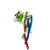
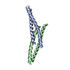
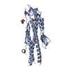
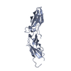
| Title | Crystal structure of Bacillus cereus FlgL |
|---|
 Components Components | Flagellar hook-associated protein 3 |
|---|
 Keywords Keywords |  STRUCTURAL PROTEIN / flagellum / junction STRUCTURAL PROTEIN / flagellum / junction |
|---|
| Function / homology | Flagellar hook-associated protein 3 / bacterial-type flagellum hook / Bacterial flagellin C-terminal helical region / Flagellin, N-terminal domain / Bacterial flagellin N-terminal helical region / bacterial-type flagellum-dependent cell motility / structural molecule activity / Flagellar hook-associated protein 3 Function and homology information Function and homology information |
|---|
| Biological species |   Bacillus cereus (bacteria) Bacillus cereus (bacteria) |
|---|
| Method | X-RAY DIFFRACTION / SYNCHROTRON / MAD / Resolution: 2.2 Å |
|---|
 Authors Authors | Hong, H.J. / Kim, T.H. / Song, W.S. / Yoon, S.I. |
|---|
 Citation Citation | Journal: Sci Rep / Year: 2018
Title: Crystal structure of FlgL and its implications for flagellar assembly
Authors: Hong, H.J. / Kim, T.H. / Song, W.S. / Ko, H.J. / Lee, G.S. / Kang, S.G. / Kim, P.H. / Yoon, S.I. |
|---|
| History | | Deposition | Mar 18, 2018 | Deposition site: PDBJ / Processing site: PDBJ |
|---|
| Revision 1.0 | Oct 17, 2018 | Provider: repository / Type: Initial release |
|---|
| Revision 1.1 | Mar 27, 2024 | Group: Data collection / Database references ...Data collection / Database references / Derived calculations / Refinement description
Category: chem_comp_atom / chem_comp_bond ...chem_comp_atom / chem_comp_bond / database_2 / pdbx_struct_conn_angle / struct_conn / struct_ncs_dom_lim
Item: _database_2.pdbx_DOI / _database_2.pdbx_database_accession ..._database_2.pdbx_DOI / _database_2.pdbx_database_accession / _pdbx_struct_conn_angle.ptnr1_auth_asym_id / _pdbx_struct_conn_angle.ptnr1_auth_comp_id / _pdbx_struct_conn_angle.ptnr1_auth_seq_id / _pdbx_struct_conn_angle.ptnr1_label_asym_id / _pdbx_struct_conn_angle.ptnr1_label_atom_id / _pdbx_struct_conn_angle.ptnr1_label_comp_id / _pdbx_struct_conn_angle.ptnr1_label_seq_id / _pdbx_struct_conn_angle.ptnr1_symmetry / _pdbx_struct_conn_angle.ptnr2_auth_seq_id / _pdbx_struct_conn_angle.ptnr2_label_asym_id / _pdbx_struct_conn_angle.ptnr2_symmetry / _pdbx_struct_conn_angle.ptnr3_auth_asym_id / _pdbx_struct_conn_angle.ptnr3_auth_comp_id / _pdbx_struct_conn_angle.ptnr3_auth_seq_id / _pdbx_struct_conn_angle.ptnr3_label_asym_id / _pdbx_struct_conn_angle.ptnr3_label_atom_id / _pdbx_struct_conn_angle.ptnr3_label_comp_id / _pdbx_struct_conn_angle.ptnr3_label_seq_id / _pdbx_struct_conn_angle.ptnr3_symmetry / _pdbx_struct_conn_angle.value / _struct_conn.pdbx_dist_value / _struct_conn.ptnr1_auth_asym_id / _struct_conn.ptnr1_auth_comp_id / _struct_conn.ptnr1_auth_seq_id / _struct_conn.ptnr1_label_asym_id / _struct_conn.ptnr1_label_atom_id / _struct_conn.ptnr1_label_comp_id / _struct_conn.ptnr1_label_seq_id / _struct_conn.ptnr2_auth_asym_id / _struct_conn.ptnr2_auth_comp_id / _struct_conn.ptnr2_auth_seq_id / _struct_conn.ptnr2_label_asym_id / _struct_conn.ptnr2_label_atom_id / _struct_conn.ptnr2_label_comp_id / _struct_conn.ptnr2_label_seq_id / _struct_conn.ptnr2_symmetry / _struct_ncs_dom_lim.beg_auth_comp_id / _struct_ncs_dom_lim.beg_label_asym_id / _struct_ncs_dom_lim.beg_label_comp_id / _struct_ncs_dom_lim.beg_label_seq_id / _struct_ncs_dom_lim.end_auth_comp_id / _struct_ncs_dom_lim.end_label_asym_id / _struct_ncs_dom_lim.end_label_comp_id / _struct_ncs_dom_lim.end_label_seq_id |
|---|
|
|---|
 Movie
Movie Controller


 Open data
 Basic information
 Structure visualization
 Downloads & links
 Other downloads


 PDBj
PDBj Assembly
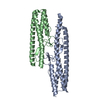


 Mass: 65.409 Da / Num. of mol.: 7 / Source method: obtained synthetically / Formula: Zn
 Mass: 18.015 Da / Num. of mol.: 88 / Source method: isolated from a natural source / Formula: H2O
 Sample preparation
 / Beamline: 7A (6B, 6C1) / Wavelength: 1.00004 Å
 Processing