 Movie
Movie Controller


+ Open data

- Basic information
| Entry | Database: PDB / ID: 5zi9 | ||||||
|---|---|---|---|---|---|---|---|
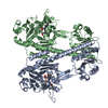
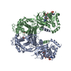
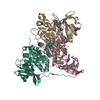
| Title | Crystal structure of type-II LOG from Streptomyces coelicolor A3 | ||||||
 Components Components | Cytokinin riboside 5'-monophosphate phosphoribohydrolase | ||||||
 Keywords Keywords |  HYDROLASE HYDROLASE | ||||||
| Function / homology | cytokinin riboside 5'-monophosphate phosphoribohydrolase activity / : / Cytokinin riboside 5'-monophosphate phosphoribohydrolase LOG / cytokinin biosynthetic process / LOG family / Possible lysine decarboxylase / cytosol / CITRATE ANION / Cytokinin riboside 5'-monophosphate phosphoribohydrolase Function and homology information Function and homology information | ||||||
| Biological species |  Streptomyces coelicolor (bacteria) Streptomyces coelicolor (bacteria) | ||||||
| Method | X-RAY DIFFRACTION / SYNCHROTRON / MOLECULAR REPLACEMENT / Resolution: 2.5 Å | ||||||
 Authors Authors | Seo, H. / Kim, K.-J. | ||||||
 Citation Citation | Journal: Biochem. Biophys. Res. Commun. / Year: 2018 Title: Structural and biochemical characterization of the type-II LOG protein from Streptomyces coelicolor A3. Authors: Seo, H. / Kim, K.J. | ||||||
| History |
|
- Structure visualization
| Structure viewer | Molecule: MolmilJmol/JSmol |
|---|
- Downloads & links
-Download
| PDBx/mmCIF format | 5zi9.cif.gz | 184.8 KB | Display | PDBx/mmCIF format |
|---|---|---|---|---|
| PDB format | pdb5zi9.ent.gz | 148.8 KB | Display | PDB format |
| PDBx/mmJSON format | 5zi9.json.gz | Tree view | PDBx/mmJSON format | |
| Others |  Other downloads Other downloads |
-Validation report
| Arichive directory | https://data.pdbj.org/pub/pdb/validation_reports/zi/5zi9ftp://data.pdbj.org/pub/pdb/validation_reports/zi/5zi9 | HTTPS FTP |
|---|
-Related structure data
| Related structure data |  5wq3S S: Starting model for refinement |
|---|---|
| Similar structure data |


-Links
 PDBj
PDBj


- Assembly
| Deposited unit | 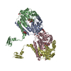
| ||||||||
|---|---|---|---|---|---|---|---|---|---|
| 1 | 
| ||||||||
| 2 | 
| ||||||||
| Unit cell |
| ||||||||
| Components on special symmetry positions |
|
-Components
| #1: Protein | Mass: 28552.422 Da / Num. of mol.: 4 Source method: isolated from a genetically manipulated source Source: (gene. exp.) Streptomyces coelicolor (strain ATCC BAA-471 / A3(2) / M145) (bacteria)Strain: ATCC BAA-471 / A3(2) / M145 / Gene: SCO5140 / Plasmid: pET30a / Production host: Escherichia coli BL21(DE3) (bacteria) / Strain (production host): BL21(DE3)References: UniProt: Q9FBL8, Hydrolases; Glycosylases; Hydrolysing N-glycosyl compounds#2: Chemical | ChemComp-EDO / Ethylene glycol  Mass: 62.068 Da / Num. of mol.: 8 / Source method: obtained synthetically / Formula: C2H6O2 Mass: 62.068 Da / Num. of mol.: 8 / Source method: obtained synthetically / Formula: C2H6O2#3: Chemical | ChemComp-FLC / Citric acid  Mass: 189.100 Da / Num. of mol.: 6 / Source method: obtained synthetically / Formula: C6H5O7 Mass: 189.100 Da / Num. of mol.: 6 / Source method: obtained synthetically / Formula: C6H5O7#4: Chemical | ChemComp-GOL / Glycerol  Mass: 92.094 Da / Num. of mol.: 4 / Source method: obtained synthetically / Formula: C3H8O3 Mass: 92.094 Da / Num. of mol.: 4 / Source method: obtained synthetically / Formula: C3H8O3#5: Water | ChemComp-HOH / | Water Mass: 18.015 Da / Num. of mol.: 69 / Source method: isolated from a natural source / Formula: H2O Mass: 18.015 Da / Num. of mol.: 69 / Source method: isolated from a natural source / Formula: H2O |
|---|
-Experimental details
-Experiment
| Experiment | Method: X-RAY DIFFRACTION / Number of used crystals: 1 |
|---|
- Sample preparation
| Crystal | Density Matthews: 3.23 Å3/Da / Density % sol: 61.86 % |
|---|---|
| Crystal grow | Temperature: 293 K / Method: vapor diffusion, hanging drop / pH: 5.5 / Details: Polyethylene glycol 3350, citrate |
-Data collection
| Diffraction | Mean temperature: 100 K |
|---|---|
| Diffraction source | Source: SYNCHROTRON / Site: PAL/PLS  / Beamline: 7A (6B, 6C1) / Wavelength: 0.97934 Å / Beamline: 7A (6B, 6C1) / Wavelength: 0.97934 Å |
| Detector | Type: ADSC QUANTUM 270 / Detector: CCD / Date: Sep 27, 2016 |
| Radiation | Monochromator: Double Crystal Monochromator / Protocol: SINGLE WAVELENGTH / Monochromatic (M) / Laue (L): M / Scattering type: x-ray |
| Radiation wavelength | Wavelength: 0.97934 Å / Relative weight: 1 |
| Reflection | Resolution: 2.5→146.21 Å / Num. obs: 49440 / % possible obs: 98 % / Redundancy: 8.7 % / Rmerge(I) obs: 0.108 / Net I/σ(I): 17.67 |
| Reflection shell | Resolution: 2.5→2.54 Å / Rmerge(I) obs: 0.31 / Mean I/σ(I) obs: 3.8 / Num. unique obs: 2396 / CC1/2: 0.374 / % possible all: 96.8 |
- Processing
| Software |
| ||||||||||||||||||||||||||||||||||||||||||||||||||||||||||||
|---|---|---|---|---|---|---|---|---|---|---|---|---|---|---|---|---|---|---|---|---|---|---|---|---|---|---|---|---|---|---|---|---|---|---|---|---|---|---|---|---|---|---|---|---|---|---|---|---|---|---|---|---|---|---|---|---|---|---|---|---|---|
| Refinement | Method to determine structure: MOLECULAR REPLACEMENT Starting model: 5WQ3 Resolution: 2.5→146.21 Å / Cor.coef. Fo:Fc: 0.942 / Cor.coef. Fo:Fc free: 0.907 / SU B: 8.666 / SU ML: 0.183 / Cross valid method: THROUGHOUT / σ(F): 0 / ESU R: 0.295 / ESU R Free: 0.237 / Stereochemistry target values: MAXIMUM LIKELIHOOD Details: HYDROGENS HAVE BEEN ADDED IN THE RIDING POSITIONS U VALUES : REFINED INDIVIDUALLY
| ||||||||||||||||||||||||||||||||||||||||||||||||||||||||||||
| Solvent computation | Ion probe radii: 0.8 Å / Shrinkage radii: 0.8 Å / VDW probe radii: 1.2 Å / Solvent model: MASK | ||||||||||||||||||||||||||||||||||||||||||||||||||||||||||||
| Displacement parameters | Biso max: 89.64 Å2 / Biso mean: 32.075 Å2 / Biso min: 16.37 Å2
| ||||||||||||||||||||||||||||||||||||||||||||||||||||||||||||
| Refinement step | Cycle: final / Resolution: 2.5→146.21 Å
| ||||||||||||||||||||||||||||||||||||||||||||||||||||||||||||
| Refine LS restraints |
| ||||||||||||||||||||||||||||||||||||||||||||||||||||||||||||
| LS refinement shell | Resolution: 2.505→2.57 Å / Rfactor Rfree error: 0 / Total num. of bins used: 20
|
