 Movie
Movie Controller


+ Open data

- Basic information
| Entry | Database: PDB / ID: 5zgn | ||||||
|---|---|---|---|---|---|---|---|
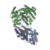
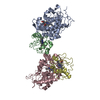
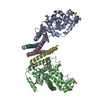
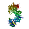
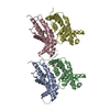
| Title | The crystal structure of KacTA-DNA complex | ||||||
 Components Components |
| ||||||
 Keywords Keywords |  TOXIN / KacT / KacA / Complex TOXIN / KacT / KacA / Complex | ||||||
| Function / homology |  Function and homology informationN-acetyltransferase activity / regulation of DNA-templated transcription Function and homology informationN-acetyltransferase activity / regulation of DNA-templated transcriptionSimilarity search - Function | ||||||
| Biological species |  Klebsiella pneumoniae subsp. pneumoniae HS11286 (bacteria) Klebsiella pneumoniae subsp. pneumoniae HS11286 (bacteria)synthetic construct (others) | ||||||
| Method | X-RAY DIFFRACTION / SYNCHROTRON / MOLECULAR REPLACEMENT / Resolution: 2.24 Å | ||||||
 Authors Authors | Qian, H.L. / Yao, Q.Q. / Gan, J.H. / Ou, H.Y. | ||||||
| Funding support |  China, 1items China, 1items
| ||||||
 Citation Citation | Journal: To Be Published Title: The crystal structure of KacTA-DNA complex Authors: Qian, H.L. / Yao, Q.Q. / Gan, J.H. / Ou, H.Y. | ||||||
| History |
|
- Structure visualization
| Structure viewer | Molecule: MolmilJmol/JSmol |
|---|
- Downloads & links
-Download
| PDBx/mmCIF format | 5zgn.cif.gz | 308.4 KB | Display | PDBx/mmCIF format |
|---|---|---|---|---|
| PDB format | pdb5zgn.ent.gz | 258.5 KB | Display | PDB format |
| PDBx/mmJSON format | 5zgn.json.gz | Tree view | PDBx/mmJSON format | |
| Others |  Other downloads Other downloads |
-Validation report
| Arichive directory | https://data.pdbj.org/pub/pdb/validation_reports/zg/5zgnftp://data.pdbj.org/pub/pdb/validation_reports/zg/5zgn | HTTPS FTP |
|---|
-Related structure data
| Similar structure data |
|---|


-Links
 PDBj
PDBj


- Assembly
| Deposited unit | 
| ||||||||
|---|---|---|---|---|---|---|---|---|---|
| 1 |
| ||||||||
| Unit cell |
|
-Components
-Protein , 2 types, 6 molecules ABDECF
| #1: Protein | Mass: 10152.146 Da / Num. of mol.: 4 Source method: isolated from a genetically manipulated source Details: KacA Source: (gene. exp.) Klebsiella pneumoniae subsp. pneumoniae HS11286 (bacteria)Strain: HS11286 / Gene: KPHS_05880 Production host: References: UniProt: A0A0H3GLZ1 #2: Protein | Mass: 20093.129 Da / Num. of mol.: 2 Source method: isolated from a genetically manipulated source Source: (gene. exp.) Klebsiella pneumoniae subsp. pneumoniae HS11286 (bacteria)Strain: HS11286 / Gene: KPHS_05890 Production host: References: UniProt: A0A0H3GMP0 |
|---|
-DNA chain , 2 types, 2 molecules GH
| #3: DNA chain | Mass: 8323.408 Da / Num. of mol.: 1 / Source method: obtained synthetically / Source: (synth.) synthetic construct (others) |
|---|---|
| #4: DNA chain | Mass: 8265.356 Da / Num. of mol.: 1 / Source method: obtained synthetically / Source: (synth.) synthetic construct (others) |
-Non-polymers , 2 types, 54 molecules 

| #5: Chemical | Sulfate Mass: 96.063 Da / Num. of mol.: 3 / Source method: obtained synthetically / Formula: SO4 Mass: 96.063 Da / Num. of mol.: 3 / Source method: obtained synthetically / Formula: SO4#6: Water | ChemComp-HOH / | WaterMass: 18.015 Da / Num. of mol.: 51 / Source method: isolated from a natural source / Formula: H2O |
|---|
-Experimental details
-Experiment
| Experiment | Method: X-RAY DIFFRACTION / Number of used crystals: 1 |
|---|
- Sample preparation
| Crystal | Density Matthews: 2.24 Å3/Da / Density % sol: 45.06 % / Description: rod |
|---|---|
| Crystal grow | Temperature: 291 K / Method: vapor diffusion, hanging drop / pH: 6.5 Details: 0.05M ammonium sulfate, 0.05M BIS-TRIS pH 6.5, 30% pentaerythritol ethoxylate (15/4 EO/OH) |
-Data collection
| Diffraction | Mean temperature: 100 K |
|---|---|
| Diffraction source | Source: SYNCHROTRON / Site: SSRF / Beamline: BL19U1 / Wavelength: 0.979 Å |
| Detector | Type: DECTRIS PILATUS3 S 6M / Detector: PIXEL / Date: Dec 20, 2017 |
| Radiation | Protocol: SINGLE WAVELENGTH / Monochromatic (M) / Laue (L): M / Scattering type: x-ray |
| Radiation wavelength | Wavelength: 0.979 Å / Relative weight: 1 |
| Reflection | Resolution: 2.24→30 Å / Num. obs: 38226 / % possible obs: 94.2 % / Redundancy: 3 % / Rmerge(I) obs: 0.076 / Net I/σ(I): 19.4 |
| Reflection shell | Resolution: 2.24→2.33 Å / Redundancy: 2.5 % / Rmerge(I) obs: 0.311 / % possible all: 92.1 |
- Processing
| Software |
| ||||||||||||||||||||||||||||||||||||||||||||||||||||||||||||||||||||||||||||||||||||||||||||||||||
|---|---|---|---|---|---|---|---|---|---|---|---|---|---|---|---|---|---|---|---|---|---|---|---|---|---|---|---|---|---|---|---|---|---|---|---|---|---|---|---|---|---|---|---|---|---|---|---|---|---|---|---|---|---|---|---|---|---|---|---|---|---|---|---|---|---|---|---|---|---|---|---|---|---|---|---|---|---|---|---|---|---|---|---|---|---|---|---|---|---|---|---|---|---|---|---|---|---|---|---|
| Refinement | Method to determine structure: MOLECULAR REPLACEMENT / Resolution: 2.24→28.44 Å / SU ML: 0.24 / Cross valid method: FREE R-VALUE / σ(F): 2.05 / Phase error: 28.35 Details: SF FILE CONTAINS FRIEDEL PAIRS UNDER I/F_MINUS AND I/F_PLUS COLUMNS.
| ||||||||||||||||||||||||||||||||||||||||||||||||||||||||||||||||||||||||||||||||||||||||||||||||||
| Solvent computation | Shrinkage radii: 0.9 Å / VDW probe radii: 1.11 Å | ||||||||||||||||||||||||||||||||||||||||||||||||||||||||||||||||||||||||||||||||||||||||||||||||||
| Refinement step | Cycle: LAST / Resolution: 2.24→28.44 Å
| ||||||||||||||||||||||||||||||||||||||||||||||||||||||||||||||||||||||||||||||||||||||||||||||||||
| Refine LS restraints |
| ||||||||||||||||||||||||||||||||||||||||||||||||||||||||||||||||||||||||||||||||||||||||||||||||||
| LS refinement shell |
| ||||||||||||||||||||||||||||||||||||||||||||||||||||||||||||||||||||||||||||||||||||||||||||||||||
| Refinement TLS params. | Method: refined / Origin x: 56.1852 Å / Origin y: -10.6855 Å / Origin z: 65.3312 Å
| ||||||||||||||||||||||||||||||||||||||||||||||||||||||||||||||||||||||||||||||||||||||||||||||||||
| Refinement TLS group | Selection details: ALL |
