 Movie
Movie Controller

[English] 日本語
 Yorodumi
- PDB-5zer: UDP Glucose alpha tetrahydrobiopterin glycosyltransferase from Sy... -
+ Open data

- Basic information
| Entry | Database: PDB / ID: 5zer | ||||||
|---|---|---|---|---|---|---|---|
| Title | UDP Glucose alpha tetrahydrobiopterin glycosyltransferase from Synechococcus species PCC 7942 - BH2 complex form | ||||||
 Components Components | UDP-glucose:tetrahydrobiopterin glucosyltransferase | ||||||
 Keywords Keywords |  TRANSFERASE / Tetrahydrobiopterin / Pteridine glycosyltransferase / Pteridine glycosides TRANSFERASE / Tetrahydrobiopterin / Pteridine glycosyltransferase / Pteridine glycosides | ||||||
| Function / homology | Glycosyl transferase, family 1 / Glycosyl transferases group 1 / glycosyltransferase activity / nucleotide binding / 7,8-DIHYDROBIOPTERIN / UDP-glucose:tetrahydrobiopterin glucosyltransferase / UDP-glucose:tetrahydrobiopterin glucosyltransferase Function and homology information Function and homology information | ||||||
| Biological species |  Synechococcus elongatus PCC 7942 (bacteria) Synechococcus elongatus PCC 7942 (bacteria) | ||||||
| Method | X-RAY DIFFRACTION / SYNCHROTRON / MOLECULAR REPLACEMENT / Resolution: 2.39 Å | ||||||
 Authors Authors | Killivalavan, A. / Lee, K.H. | ||||||
| Funding support |  Korea, Republic Of, 1items Korea, Republic Of, 1items
| ||||||
 Citation Citation | Journal: To Be Published Title: UDP Glucose alpha tetrahydrobiopterin glycosyltransferase from Synechococcus species PCC 7942 - BH2 complex form Authors: Killivalavan, A. / Lee, K.H. | ||||||
| History |
|
- Structure visualization
| Structure viewer | Molecule: MolmilJmol/JSmol |
|---|
- Downloads & links
-Download
| PDBx/mmCIF format | 5zer.cif.gz | 150.1 KB | Display | PDBx/mmCIF format |
|---|---|---|---|---|
| PDB format | pdb5zer.ent.gz | 117.4 KB | Display | PDB format |
| PDBx/mmJSON format | 5zer.json.gz | Tree view | PDBx/mmJSON format | |
| Others |  Other downloads Other downloads |
-Validation report
| Arichive directory | https://data.pdbj.org/pub/pdb/validation_reports/ze/5zerftp://data.pdbj.org/pub/pdb/validation_reports/ze/5zer | HTTPS FTP |
|---|
-Related structure data
| Related structure data |  5ze7S S: Starting model for refinement |
|---|---|
| Similar structure data |


-Links
 PDBj
PDBj


- Assembly
| Deposited unit | 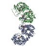
| ||||||||
|---|---|---|---|---|---|---|---|---|---|
| 1 |
| ||||||||
| Unit cell |
|
-Components
| #1: Protein | Mass: 38406.992 Da / Num. of mol.: 2 / Fragment: UNP residues 2-355 Source method: isolated from a genetically manipulated source Source: (gene. exp.) Synechococcus elongatus PCC 7942 (bacteria)Strain: PCC 7942 / Production host: Escherichia coli BL21(DE3) (bacteria) / Strain (production host): BL21(DE3) / References: UniProt: Q93EY3, UniProt: Q31LX1*PLUS#2: Chemical | Dihydrobiopterin  Mass: 239.231 Da / Num. of mol.: 2 / Source method: obtained synthetically / Formula: C9H13N5O3 / Feature type: SUBJECT OF INVESTIGATION Mass: 239.231 Da / Num. of mol.: 2 / Source method: obtained synthetically / Formula: C9H13N5O3 / Feature type: SUBJECT OF INVESTIGATION#3: Chemical | ChemComp-GOL / | Glycerol  Mass: 92.094 Da / Num. of mol.: 1 / Source method: obtained synthetically / Formula: C3H8O3 Mass: 92.094 Da / Num. of mol.: 1 / Source method: obtained synthetically / Formula: C3H8O3#4: Water | ChemComp-HOH / | Water Mass: 18.015 Da / Num. of mol.: 159 / Source method: isolated from a natural source / Formula: H2O Mass: 18.015 Da / Num. of mol.: 159 / Source method: isolated from a natural source / Formula: H2O |
|---|
-Experimental details
-Experiment
| Experiment | Method: X-RAY DIFFRACTION / Number of used crystals: 1 |
|---|
- Sample preparation
| Crystal | Density Matthews: 2.32 Å3/Da / Density % sol: 47.03 % |
|---|---|
| Crystal grow | Temperature: 291 K / Method: vapor diffusion, hanging drop / Details: Bis-tris, PEG3350, Galactose |
-Data collection
| Diffraction | Mean temperature: 100 K |
|---|---|
| Diffraction source | Source: SYNCHROTRON / Site: Photon Factory  / Beamline: BL-17A / Wavelength: 0.98 Å / Beamline: BL-17A / Wavelength: 0.98 Å |
| Detector | Type: ADSC QUANTUM 270 / Detector: CCD / Date: May 28, 2013 |
| Radiation | Protocol: SINGLE WAVELENGTH / Monochromatic (M) / Laue (L): M / Scattering type: x-ray |
| Radiation wavelength | Wavelength: 0.98 Å / Relative weight: 1 |
| Reflection | Resolution: 2.39→50 Å / Num. obs: 26482 / % possible obs: 98.4 % / Redundancy: 4.1 % / Net I/σ(I): 8.4 |
| Reflection shell | Resolution: 2.59→2.63 Å |
- Processing
| Software |
| ||||||||||||||||||||||||||||||||||||||||||||||||||||||||||||||||||||||||||||||||||||||||||||||||||||||||||||||||||||||||||||||||||||||||||||||||||||||||||||||||||||||||||||||||||||||
|---|---|---|---|---|---|---|---|---|---|---|---|---|---|---|---|---|---|---|---|---|---|---|---|---|---|---|---|---|---|---|---|---|---|---|---|---|---|---|---|---|---|---|---|---|---|---|---|---|---|---|---|---|---|---|---|---|---|---|---|---|---|---|---|---|---|---|---|---|---|---|---|---|---|---|---|---|---|---|---|---|---|---|---|---|---|---|---|---|---|---|---|---|---|---|---|---|---|---|---|---|---|---|---|---|---|---|---|---|---|---|---|---|---|---|---|---|---|---|---|---|---|---|---|---|---|---|---|---|---|---|---|---|---|---|---|---|---|---|---|---|---|---|---|---|---|---|---|---|---|---|---|---|---|---|---|---|---|---|---|---|---|---|---|---|---|---|---|---|---|---|---|---|---|---|---|---|---|---|---|---|---|---|---|
| Refinement | Method to determine structure: MOLECULAR REPLACEMENT Starting model: 5ZE7 Resolution: 2.39→45.71 Å / Cor.coef. Fo:Fc: 0.936 / Cor.coef. Fo:Fc free: 0.886 / SU B: 14.89 / SU ML: 0.315 / Cross valid method: THROUGHOUT / ESU R: 0.588 / ESU R Free: 0.318 / Details: HYDROGENS HAVE BEEN ADDED IN THE RIDING POSITIONS
| ||||||||||||||||||||||||||||||||||||||||||||||||||||||||||||||||||||||||||||||||||||||||||||||||||||||||||||||||||||||||||||||||||||||||||||||||||||||||||||||||||||||||||||||||||||||
| Solvent computation | Ion probe radii: 0.8 Å / Shrinkage radii: 0.8 Å / VDW probe radii: 1.2 Å | ||||||||||||||||||||||||||||||||||||||||||||||||||||||||||||||||||||||||||||||||||||||||||||||||||||||||||||||||||||||||||||||||||||||||||||||||||||||||||||||||||||||||||||||||||||||
| Displacement parameters | Biso mean: 50.664 Å2
| ||||||||||||||||||||||||||||||||||||||||||||||||||||||||||||||||||||||||||||||||||||||||||||||||||||||||||||||||||||||||||||||||||||||||||||||||||||||||||||||||||||||||||||||||||||||
| Refinement step | Cycle: 1 / Resolution: 2.39→45.71 Å
| ||||||||||||||||||||||||||||||||||||||||||||||||||||||||||||||||||||||||||||||||||||||||||||||||||||||||||||||||||||||||||||||||||||||||||||||||||||||||||||||||||||||||||||||||||||||
| Refine LS restraints |
|