 Movie
Movie Controller

[English] 日本語
 Yorodumi
- PDB-5zdb: Crystal structure of poly(ADP-ribose) glycohydrolase (PARG) from ... -
+ Open data

- Basic information
| Entry | Database: PDB / ID: 5zdb | ||||||
|---|---|---|---|---|---|---|---|
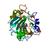
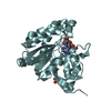
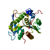
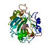
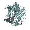
| Title | Crystal structure of poly(ADP-ribose) glycohydrolase (PARG) from Deinococcus radiodurans in complex with ADP-ribose (P21) | ||||||
 Components Components | Poly ADP-ribose glycohydrolase | ||||||
 Keywords Keywords |  HYDROLASE / ADP-ribose / poly(ADP-ribose) glycohydrolase HYDROLASE / ADP-ribose / poly(ADP-ribose) glycohydrolase | ||||||
| Function / homology |  Function and homology information Function and homology informationConserved hypothetical protein CHP02452 / Microbial-type PARG, catalytic domain / Microbial-type PARG, catalytic domain / Leucine Aminopeptidase, subunit E, domain 1 / Leucine Aminopeptidase, subunit E; domain 1 / Macro domain-like / 3-Layer(aba) Sandwich / Alpha BetaSimilarity search - Domain/homology | ||||||
| Biological species |  Deinococcus radiodurans (radioresistant) Deinococcus radiodurans (radioresistant) | ||||||
| Method | X-RAY DIFFRACTION / SYNCHROTRON / MOLECULAR REPLACEMENT / Resolution: 1.972 Å | ||||||
 Authors Authors | Cho, C.C. / Hsu, C.H. | ||||||
| Funding support |  Taiwan, 1items Taiwan, 1items
| ||||||
 Citation Citation | Journal: Nat Commun / Year: 2019 Title: Structural and biochemical evidence supporting poly ADP-ribosylation in the bacterium Deinococcus radiodurans. Authors: Cho, C.C. / Chien, C.Y. / Chiu, Y.C. / Lin, M.H. / Hsu, C.H. | ||||||
| History |
|
- Structure visualization
| Structure viewer | Molecule: MolmilJmol/JSmol |
|---|
- Downloads & links
-Download
| PDBx/mmCIF format | 5zdb.cif.gz | 214.5 KB | Display | PDBx/mmCIF format |
|---|---|---|---|---|
| PDB format | pdb5zdb.ent.gz | 169.9 KB | Display | PDB format |
| PDBx/mmJSON format | 5zdb.json.gz | Tree view | PDBx/mmJSON format | |
| Others |  Other downloads Other downloads |
-Validation report
| Arichive directory | https://data.pdbj.org/pub/pdb/validation_reports/zd/5zdbftp://data.pdbj.org/pub/pdb/validation_reports/zd/5zdb | HTTPS FTP |
|---|
-Related structure data
| Related structure data | 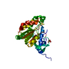 5zdaSC 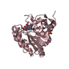 5zdcC 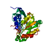 5zddC 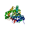 5zdeC  5zdfC 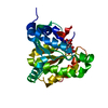 5zdgC S: Starting model for refinement C: citing same article ( |
|---|---|
| Similar structure data |


-Links
 PDBj
PDBj
- Assembly
| Deposited unit | 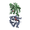
| ||||||||
|---|---|---|---|---|---|---|---|---|---|
| 1 | 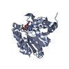
| ||||||||
| 2 | 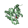
| ||||||||
| Unit cell |
|
-Components
| #1: Protein | Mass: 27600.174 Da / Num. of mol.: 2 Source method: isolated from a genetically manipulated source Source: (gene. exp.) Deinococcus radiodurans (strain ATCC 13939 / DSM 20539 / JCM 16871 / LMG 4051 / NBRC 15346 / NCIMB 9279 / R1 / VKM B-1422) (radioresistant)Strain: ATCC 13939 / DSM 20539 / JCM 16871 / LMG 4051 / NBRC 15346 / NCIMB 9279 / R1 / VKM B-1422 Gene: DR_B0099 / Production host: Escherichia coli BL21(DE3) (bacteria) / Strain (production host): BL21(DE3) / References: UniProt: Q9RZM4#2: Chemical |   Mass: 559.316 Da / Num. of mol.: 2 / Source method: obtained synthetically / Formula: C15H23N5O14P2 Mass: 559.316 Da / Num. of mol.: 2 / Source method: obtained synthetically / Formula: C15H23N5O14P2#3: Water | ChemComp-HOH / | Water Mass: 18.015 Da / Num. of mol.: 266 / Source method: isolated from a natural source / Formula: H2O Mass: 18.015 Da / Num. of mol.: 266 / Source method: isolated from a natural source / Formula: H2O |
|---|
-Experimental details
-Experiment
| Experiment | Method: X-RAY DIFFRACTION / Number of used crystals: 1 |
|---|
- Sample preparation
| Crystal | Density Matthews: 2.27 Å3/Da / Density % sol: 45.87 % |
|---|---|
| Crystal grow | Temperature: 283 K / Method: vapor diffusion, sitting drop / pH: 6 Details: 0.1 M Sodium citrate tribasic dihydrate pH 6.0, 10% 2-Propanol, 10% Polyethylene glycol 4000 |
-Data collection
| Diffraction | Mean temperature: 100 K |
|---|---|
| Diffraction source | Source: SYNCHROTRON / Site: NSRRC / Beamline: BL13C1 / Wavelength: 0.97622 Å |
| Detector | Type: ADSC QUANTUM 315r / Detector: CCD / Date: May 15, 2014 |
| Radiation | Protocol: SINGLE WAVELENGTH / Monochromatic (M) / Laue (L): M / Scattering type: x-ray |
| Radiation wavelength | Wavelength: 0.97622 Å / Relative weight: 1 |
| Reflection | Resolution: 1.972→25.959 Å / Num. obs: 33944 / % possible obs: 98.2 % / Redundancy: 3.8 % / Rmerge(I) obs: 0.086 / Net I/σ(I): 14.353 |
| Reflection shell | Resolution: 1.97→2.04 Å / Redundancy: 3.3 % / Rmerge(I) obs: 0.502 / Mean I/σ(I) obs: 2.019 / % possible all: 91.1 |
- Processing
| Software |
| ||||||||||||||||||||||||||||||||||||||||||||||||||||||||||||||||||||||||||||||||||||||||||||||||||||||||||||||||||||||||||||||||||||||||||||||||||||||
|---|---|---|---|---|---|---|---|---|---|---|---|---|---|---|---|---|---|---|---|---|---|---|---|---|---|---|---|---|---|---|---|---|---|---|---|---|---|---|---|---|---|---|---|---|---|---|---|---|---|---|---|---|---|---|---|---|---|---|---|---|---|---|---|---|---|---|---|---|---|---|---|---|---|---|---|---|---|---|---|---|---|---|---|---|---|---|---|---|---|---|---|---|---|---|---|---|---|---|---|---|---|---|---|---|---|---|---|---|---|---|---|---|---|---|---|---|---|---|---|---|---|---|---|---|---|---|---|---|---|---|---|---|---|---|---|---|---|---|---|---|---|---|---|---|---|---|---|---|---|---|---|
| Refinement | Method to determine structure: MOLECULAR REPLACEMENT Starting model: 5ZDA Resolution: 1.972→25.959 Å / SU ML: 0.21 / Cross valid method: THROUGHOUT / σ(F): 1.35 / Phase error: 19.81
| ||||||||||||||||||||||||||||||||||||||||||||||||||||||||||||||||||||||||||||||||||||||||||||||||||||||||||||||||||||||||||||||||||||||||||||||||||||||
| Solvent computation | Shrinkage radii: 0.9 Å / VDW probe radii: 1.11 Å | ||||||||||||||||||||||||||||||||||||||||||||||||||||||||||||||||||||||||||||||||||||||||||||||||||||||||||||||||||||||||||||||||||||||||||||||||||||||
| Refinement step | Cycle: LAST / Resolution: 1.972→25.959 Å
| ||||||||||||||||||||||||||||||||||||||||||||||||||||||||||||||||||||||||||||||||||||||||||||||||||||||||||||||||||||||||||||||||||||||||||||||||||||||
| Refine LS restraints |
| ||||||||||||||||||||||||||||||||||||||||||||||||||||||||||||||||||||||||||||||||||||||||||||||||||||||||||||||||||||||||||||||||||||||||||||||||||||||
| LS refinement shell |
| ||||||||||||||||||||||||||||||||||||||||||||||||||||||||||||||||||||||||||||||||||||||||||||||||||||||||||||||||||||||||||||||||||||||||||||||||||||||
| Refinement TLS params. | Method: refined / Refine-ID: X-RAY DIFFRACTION
| ||||||||||||||||||||||||||||||||||||||||||||||||||||||||||||||||||||||||||||||||||||||||||||||||||||||||||||||||||||||||||||||||||||||||||||||||||||||
| Refinement TLS group |
|
