 Movie
Movie Controller

[English] 日本語
 Yorodumi
- PDB-5z1p: Structural basis of the improved sweetness and stability of the s... -
+ Open data

- Basic information
| Entry | Database: PDB / ID: 5z1p | ||||||
|---|---|---|---|---|---|---|---|
| Title | Structural basis of the improved sweetness and stability of the single-chain sweet-tasting protein monellin (MNEI) | ||||||
 Components Components | Monellin chain B,Monellin chain A | ||||||
 Keywords Keywords |  PLANT PROTEIN / monellin PLANT PROTEIN / monellin | ||||||
| Function / homology | Monellin, A chain / Monellin, A chain superfamily / Monellin, B chain / Monellin / Monellin / Cystatin superfamily / Monellin chain A / Monellin chain B Function and homology information Function and homology information | ||||||
| Biological species |  Dioscoreophyllum cumminsii (serendipity berry) Dioscoreophyllum cumminsii (serendipity berry) | ||||||
| Method | X-RAY DIFFRACTION / SYNCHROTRON / MOLECULAR REPLACEMENT / Resolution: 1.89 Å | ||||||
 Authors Authors | Liu, B. | ||||||
 Citation Citation | Journal: Biochimie / Year: 2018 Title: Structure basis of the improved sweetness and thermostability of a unique double-sites single-chain sweet-tasting protein monellin (MNEI) mutant Authors: Zhao, M. / Xu, X. / Liu, B. | ||||||
| History |
|
- Structure visualization
| Structure viewer | Molecule: MolmilJmol/JSmol |
|---|
- Downloads & links
-Download
| PDBx/mmCIF format | 5z1p.cif.gz | 93.3 KB | Display | PDBx/mmCIF format |
|---|---|---|---|---|
| PDB format | pdb5z1p.ent.gz | 70.5 KB | Display | PDB format |
| PDBx/mmJSON format | 5z1p.json.gz | Tree view | PDBx/mmJSON format | |
| Others |  Other downloads Other downloads |
-Validation report
| Arichive directory | https://data.pdbj.org/pub/pdb/validation_reports/z1/5z1pftp://data.pdbj.org/pub/pdb/validation_reports/z1/5z1p | HTTPS FTP |
|---|
-Related structure data
| Related structure data |  1iv7S S: Starting model for refinement |
|---|---|
| Similar structure data |


-Links
 PDBj
PDBj
- Assembly
| Deposited unit | 
| ||||||||
|---|---|---|---|---|---|---|---|---|---|
| 1 | 
| ||||||||
| 2 | 
| ||||||||
| 3 | 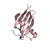
| ||||||||
| 4 | 
| ||||||||
| Unit cell |
|
-Components
| #1: Protein | Mass: 11346.002 Da / Num. of mol.: 4 / Mutation: E2N, E23A Source method: isolated from a genetically manipulated source Source: (gene. exp.) Dioscoreophyllum cumminsii (serendipity berry)Production host:  Escherichia coli (E. coli) / References: UniProt: P02882, UniProt: P02881 Escherichia coli (E. coli) / References: UniProt: P02882, UniProt: P02881#2: Water | ChemComp-HOH / | Water Mass: 18.015 Da / Num. of mol.: 277 / Source method: isolated from a natural source / Formula: H2O Mass: 18.015 Da / Num. of mol.: 277 / Source method: isolated from a natural source / Formula: H2O |
|---|
-Experimental details
-Experiment
| Experiment | Method: X-RAY DIFFRACTION / Number of used crystals: 1 |
|---|
- Sample preparation
| Crystal | Density Matthews: 2.21 Å3/Da / Density % sol: 44.39 % |
|---|---|
| Crystal grow | Temperature: 297 K / Method: vapor diffusion, sitting drop / Details: 20% PEG3350, 0.2M Sodium Aceteta, 0.1M Tris PH 8.5 |
-Data collection
| Diffraction | Mean temperature: 100 K |
|---|---|
| Diffraction source | Source: SYNCHROTRON / Site: SSRF  / Beamline: BL18U1 / Wavelength: 0.987 Å / Beamline: BL18U1 / Wavelength: 0.987 Å |
| Detector | Type: DECTRIS PILATUS3 S 6M / Detector: PIXEL / Date: Nov 1, 2017 |
| Radiation | Protocol: SINGLE WAVELENGTH / Monochromatic (M) / Laue (L): M / Scattering type: x-ray |
| Radiation wavelength | Wavelength: 0.987 Å / Relative weight: 1 |
| Reflection | Resolution: 1.9→50 Å / Num. obs: 29868 / % possible obs: 96.8 % / Redundancy: 3.3 % / Rmerge(I) obs: 0.134 / Rpim(I) all: 0.081 / Rsym value: 0.152 / Net I/σ(I): 12.75 |
| Reflection shell | Resolution: 1.9→1.93 Å / Redundancy: 2.7 % / Rmerge(I) obs: 0.418 / Num. unique obs: 1400 / Rpim(I) all: 0.252 / Rsym value: 0.452 / % possible all: 90.8 |
- Processing
| Software |
| ||||||||||||||||||||||||||||||||||||||||||||||||||||||||||||||||||||||||||||||||||||||||||||||||||||||||||||||||||||||||||||||||||||||||||||||||||||||||||||||||||||||||||||||||||||||
|---|---|---|---|---|---|---|---|---|---|---|---|---|---|---|---|---|---|---|---|---|---|---|---|---|---|---|---|---|---|---|---|---|---|---|---|---|---|---|---|---|---|---|---|---|---|---|---|---|---|---|---|---|---|---|---|---|---|---|---|---|---|---|---|---|---|---|---|---|---|---|---|---|---|---|---|---|---|---|---|---|---|---|---|---|---|---|---|---|---|---|---|---|---|---|---|---|---|---|---|---|---|---|---|---|---|---|---|---|---|---|---|---|---|---|---|---|---|---|---|---|---|---|---|---|---|---|---|---|---|---|---|---|---|---|---|---|---|---|---|---|---|---|---|---|---|---|---|---|---|---|---|---|---|---|---|---|---|---|---|---|---|---|---|---|---|---|---|---|---|---|---|---|---|---|---|---|---|---|---|---|---|---|---|
| Refinement | Method to determine structure: MOLECULAR REPLACEMENT Starting model: 1IV7 Resolution: 1.89→40.93 Å / Cor.coef. Fo:Fc: 0.942 / Cor.coef. Fo:Fc free: 0.919 / SU B: 4.503 / SU ML: 0.134 / Cross valid method: THROUGHOUT / ESU R: 0.198 / ESU R Free: 0.178 / Stereochemistry target values: MAXIMUM LIKELIHOOD / Details: HYDROGENS HAVE BEEN ADDED IN THE RIDING POSITIONS
| ||||||||||||||||||||||||||||||||||||||||||||||||||||||||||||||||||||||||||||||||||||||||||||||||||||||||||||||||||||||||||||||||||||||||||||||||||||||||||||||||||||||||||||||||||||||
| Solvent computation | Ion probe radii: 0.8 Å / Shrinkage radii: 0.8 Å / VDW probe radii: 1.2 Å / Solvent model: MASK | ||||||||||||||||||||||||||||||||||||||||||||||||||||||||||||||||||||||||||||||||||||||||||||||||||||||||||||||||||||||||||||||||||||||||||||||||||||||||||||||||||||||||||||||||||||||
| Displacement parameters | Biso mean: 33.069 Å2
| ||||||||||||||||||||||||||||||||||||||||||||||||||||||||||||||||||||||||||||||||||||||||||||||||||||||||||||||||||||||||||||||||||||||||||||||||||||||||||||||||||||||||||||||||||||||
| Refinement step | Cycle: 1 / Resolution: 1.89→40.93 Å
| ||||||||||||||||||||||||||||||||||||||||||||||||||||||||||||||||||||||||||||||||||||||||||||||||||||||||||||||||||||||||||||||||||||||||||||||||||||||||||||||||||||||||||||||||||||||
| Refine LS restraints |
|