 Movie
Movie Controller

[English] 日本語
 Yorodumi
- PDB-5yy5: Structural definition of a unique neutralization epitope on the r... -
+ Open data

- Basic information
| Entry | Database: PDB / ID: 5yy5 | ||||||
|---|---|---|---|---|---|---|---|
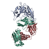
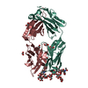
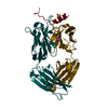
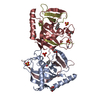
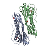
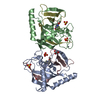
| Title | Structural definition of a unique neutralization epitope on the receptor-binding domain of MERS-CoV spike glycoprotein | ||||||
 Components Components |
| ||||||
 Keywords Keywords |  VIRAL PROTEIN / MERS-CoV / spike glycorptotein / neutralizing antibody VIRAL PROTEIN / MERS-CoV / spike glycorptotein / neutralizing antibody | ||||||
| Function / homology |  Function and homology information Function and homology informationendocytosis involved in viral entry into host cell / host cell endoplasmic reticulum-Golgi intermediate compartment membrane / membrane fusion / positive regulation of viral entry into host cell / receptor-mediated virion attachment to host cell / fusion of virus membrane with host plasma membrane / fusion of virus membrane with host endosome membrane / viral envelope / host cell plasma membrane / virion membrane / membraneSimilarity search - Function | ||||||
| Biological species |  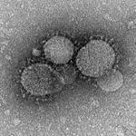 Middle East respiratory syndrome coronavirus Middle East respiratory syndrome coronavirus Homo sapiens (human) Homo sapiens (human) | ||||||
| Method | X-RAY DIFFRACTION / SYNCHROTRON / MOLECULAR REPLACEMENT / Resolution: 2.8 Å | ||||||
 Authors Authors | Zhang, S. / Wang, P. / Zhou, P. / Wang, X. / Zhang, L. | ||||||
 Citation Citation | Journal: Cell Rep / Year: 2018 Title: Structural Definition of a Unique Neutralization Epitope on the Receptor-Binding Domain of MERS-CoV Spike Glycoprotein Authors: Zhang, S. / Zhou, P. / Wang, P. / Li, Y. / Jiang, L. / Jia, W. / Wang, H. / Fan, A. / Wang, D. / Shi, X. / Fang, X. / Hammel, M. / Wang, S. / Wang, X. / Zhang, L. | ||||||
| History |
|
- Structure visualization
| Structure viewer | Molecule: MolmilJmol/JSmol |
|---|
- Downloads & links
-Download
| PDBx/mmCIF format | 5yy5.cif.gz | 336.5 KB | Display | PDBx/mmCIF format |
|---|---|---|---|---|
| PDB format | pdb5yy5.ent.gz | 282.2 KB | Display | PDB format |
| PDBx/mmJSON format | 5yy5.json.gz | Tree view | PDBx/mmJSON format | |
| Others |  Other downloads Other downloads |
-Validation report
| Arichive directory | https://data.pdbj.org/pub/pdb/validation_reports/yy/5yy5ftp://data.pdbj.org/pub/pdb/validation_reports/yy/5yy5 | HTTPS FTP |
|---|
-Related structure data


-Links
 PDBj
PDBj


- Assembly
| Deposited unit | 
| ||||||||
|---|---|---|---|---|---|---|---|---|---|
| 1 | 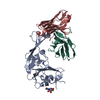
| ||||||||
| 2 | 
| ||||||||
| Unit cell |
|
-Components
-Antibody , 4 types, 4 molecules HLCD
| #2: Antibody | Mass: 12474.881 Da / Num. of mol.: 1 Source method: isolated from a genetically manipulated source Source: (gene. exp.) Homo sapiens (human) / Cell line (production host): HEK293 / Production host: Homo sapiens (human) |
|---|---|
| #3: Antibody | Mass: 11720.712 Da / Num. of mol.: 1 Source method: isolated from a genetically manipulated source Source: (gene. exp.) Homo sapiens (human) / Production host: Homo sapiens (human) |
| #4: Antibody | Mass: 12216.610 Da / Num. of mol.: 1 Source method: isolated from a genetically manipulated source Source: (gene. exp.) Homo sapiens (human) / Production host: Homo sapiens (human) |
| #5: Antibody | Mass: 11039.918 Da / Num. of mol.: 1 Source method: isolated from a genetically manipulated source Source: (gene. exp.) Homo sapiens (human) / Production host: Homo sapiens (human) |
-Protein / Sugars , 2 types, 4 molecules AB
| #1: Protein | Mass: 22987.012 Da / Num. of mol.: 2 Source method: isolated from a genetically manipulated source Source: (gene. exp.) Middle East respiratory syndrome coronavirusProduction host:   Spodoptera frugiperda (fall armyworm) / References: UniProt: K9N5Q8*PLUS Spodoptera frugiperda (fall armyworm) / References: UniProt: K9N5Q8*PLUS#6: Sugar | N-Acetylglucosamine Type: D-saccharide, beta linking / Mass: 221.208 Da / Num. of mol.: 2 Type: D-saccharide, beta linking / Mass: 221.208 Da / Num. of mol.: 2Source method: isolated from a genetically manipulated source Formula: C8H15NO6 |
|---|
-Experimental details
-Experiment
| Experiment | Method: X-RAY DIFFRACTION / Number of used crystals: 1 |
|---|
- Sample preparation
| Crystal | Density Matthews: 3.5 Å3/Da / Density % sol: 64.9 % |
|---|---|
| Crystal grow | Temperature: 291.15 K / Method: vapor diffusion / pH: 7 Details: tris pH7.5, polyethylene glycol 8000, ethylene glycol |
-Data collection
| Diffraction | Mean temperature: 100 K |
|---|---|
| Diffraction source | Source: SYNCHROTRON / Site: SSRF  / Beamline: BL17U1 / Wavelength: 0.987 Å / Beamline: BL17U1 / Wavelength: 0.987 Å |
| Detector | Type: ADSC QUANTUM 315r / Detector: CCD / Date: Jul 5, 2017 |
| Radiation | Protocol: SINGLE WAVELENGTH / Monochromatic (M) / Laue (L): M / Scattering type: x-ray |
| Radiation wavelength | Wavelength: 0.987 Å / Relative weight: 1 |
| Reflection | Resolution: 2.8→50 Å / Num. obs: 31541 / % possible obs: 99.2 % / Redundancy: 5.5 % / Net I/σ(I): 16.2 |
| Reflection shell | Resolution: 2.8→2.88 Å / Num. unique obs: 31541 / % possible all: 99.7 |
- Processing
| Software |
| ||||||||||||||||||||||||||||||||||||||||||||||||||||||||||||||||||||||||||||||||||||
|---|---|---|---|---|---|---|---|---|---|---|---|---|---|---|---|---|---|---|---|---|---|---|---|---|---|---|---|---|---|---|---|---|---|---|---|---|---|---|---|---|---|---|---|---|---|---|---|---|---|---|---|---|---|---|---|---|---|---|---|---|---|---|---|---|---|---|---|---|---|---|---|---|---|---|---|---|---|---|---|---|---|---|---|---|---|
| Refinement | Method to determine structure: MOLECULAR REPLACEMENT / Resolution: 2.8→41.51 Å / SU ML: 0.44 / Cross valid method: FREE R-VALUE / σ(F): 1.97 / Phase error: 40.11
| ||||||||||||||||||||||||||||||||||||||||||||||||||||||||||||||||||||||||||||||||||||
| Solvent computation | Shrinkage radii: 0.9 Å / VDW probe radii: 1.11 Å | ||||||||||||||||||||||||||||||||||||||||||||||||||||||||||||||||||||||||||||||||||||
| Refinement step | Cycle: LAST / Resolution: 2.8→41.51 Å
| ||||||||||||||||||||||||||||||||||||||||||||||||||||||||||||||||||||||||||||||||||||
| Refine LS restraints |
| ||||||||||||||||||||||||||||||||||||||||||||||||||||||||||||||||||||||||||||||||||||
| LS refinement shell |
| ||||||||||||||||||||||||||||||||||||||||||||||||||||||||||||||||||||||||||||||||||||
| Refinement TLS params. | Method: refined / Origin x: 4.1861 Å / Origin y: 42.723 Å / Origin z: -102.7992 Å
| ||||||||||||||||||||||||||||||||||||||||||||||||||||||||||||||||||||||||||||||||||||
| Refinement TLS group | Selection details: all |
