 Movie
Movie Controller

[English] 日本語
 Yorodumi
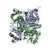
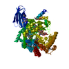
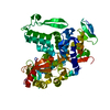
- PDB-5yx6: Crystal structure of Rv3272 from M. tuberculosis orthorhombic form -
+ Open data

- Basic information
| Entry | Database: PDB / ID: 5yx6 | |||||||||
|---|---|---|---|---|---|---|---|---|---|---|
| Title | Crystal structure of Rv3272 from M. tuberculosis orthorhombic form | |||||||||
 Components Components | Uncharacterized protein Rv3272 | |||||||||
 Keywords Keywords |  TRANSFERASE / Rv3272 / CoA transferase / fatty acid metabolism / tuberculosis TRANSFERASE / Rv3272 / CoA transferase / fatty acid metabolism / tuberculosis | |||||||||
| Function / homology | Transferases; Transferring sulfur-containing groups; CoA-transferases / CoA-transferase family III domain 3 superfamily / CoA-transferase family III / CoA-transferase family III domain 1 superfamily / CoA-transferase family III / transferase activity / BENZAMIDINE / Probable fatty acyl-CoA transferase Rv3272 Function and homology information Function and homology information | |||||||||
| Biological species |   Mycobacterium tuberculosis (bacteria) Mycobacterium tuberculosis (bacteria) | |||||||||
| Method | X-RAY DIFFRACTION / SYNCHROTRON / MOLECULAR REPLACEMENT / Resolution: 2.2 Å | |||||||||
 Authors Authors | Ansari, A. / Shrimant, K.S. / Venkatesh Pratap, J. | |||||||||
 Citation Citation | Journal: Biochim Biophys Acta Proteins Proteom / Year: 2019 Title: Rv3272 encodes a novel Family III CoA transferase that alters the cell wall lipid profile and protects mycobacteria from acidic and oxidative stress. Authors: Karade, S.S. / Pandey, S. / Ansari, A. / Das, S. / Tripathi, S. / Arora, A. / Chopra, S. / Pratap, J.V. / Dasgupta, A. | |||||||||
| History |
|
- Structure visualization
| Structure viewer | Molecule: MolmilJmol/JSmol |
|---|
- Downloads & links
-Download
| PDBx/mmCIF format | 5yx6.cif.gz | 280.5 KB | Display | PDBx/mmCIF format |
|---|---|---|---|---|
| PDB format | pdb5yx6.ent.gz | 225.6 KB | Display | PDB format |
| PDBx/mmJSON format | 5yx6.json.gz | Tree view | PDBx/mmJSON format | |
| Others |  Other downloads Other downloads |
-Validation report
| Arichive directory | https://data.pdbj.org/pub/pdb/validation_reports/yx/5yx6ftp://data.pdbj.org/pub/pdb/validation_reports/yx/5yx6 | HTTPS FTP |
|---|
-Related structure data
| Related structure data |  5yitSC  5yiyC S: Starting model for refinement C: citing same article ( |
|---|---|
| Similar structure data |


-Links
 PDBj
PDBj
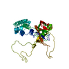
- Assembly
| Deposited unit | 
| ||||||||
|---|---|---|---|---|---|---|---|---|---|
| 1 | 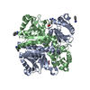
| ||||||||
| 2 | 
| ||||||||
| Unit cell |
|
-Components
| #1: Protein | Mass: 42505.445 Da / Num. of mol.: 4 Source method: isolated from a genetically manipulated source Source: (gene. exp.) Mycobacterium tuberculosis (strain ATCC 25618 / H37Rv) (bacteria)Strain: ATCC 25618 / H37Rv / Gene: Rv3272 / Production host: Escherichia coli Bl21(DE3) (bacteria) / Strain (production host): Bl21(DE3) / References: UniProt: P96877#2: Chemical | ChemComp-BEN / Benzamidine  Mass: 120.152 Da / Num. of mol.: 7 / Source method: obtained synthetically / Formula: C7H8N2 Mass: 120.152 Da / Num. of mol.: 7 / Source method: obtained synthetically / Formula: C7H8N2#3: Chemical | ChemComp-GOL / Glycerol  Mass: 92.094 Da / Num. of mol.: 6 / Source method: obtained synthetically / Formula: C3H8O3 Mass: 92.094 Da / Num. of mol.: 6 / Source method: obtained synthetically / Formula: C3H8O3#4: Water | ChemComp-HOH / | Water Mass: 18.015 Da / Num. of mol.: 348 / Source method: isolated from a natural source / Formula: H2O Mass: 18.015 Da / Num. of mol.: 348 / Source method: isolated from a natural source / Formula: H2O |
|---|
-Experimental details
-Experiment
| Experiment | Method: X-RAY DIFFRACTION / Number of used crystals: 1 |
|---|
- Sample preparation
| Crystal | Density Matthews: 2.48 Å3/Da / Density % sol: 50.34 % / Description: elongated plate-like |
|---|---|
| Crystal grow | Temperature: 279 K / Method: vapor diffusion, hanging drop / pH: 8 Details: 200mM Disodium hydrogen phosphate, 22% PEG 3350, 2% Benzamidine hydrochloride |
-Data collection
| Diffraction | Mean temperature: 100 K |
|---|---|
| Diffraction source | Source: SYNCHROTRON / Site: ESRF  / Beamline: ID30B / Wavelength: 0.966 Å / Beamline: ID30B / Wavelength: 0.966 Å |
| Detector | Type: DECTRIS PILATUS 2M / Detector: PIXEL / Date: Jul 3, 2017 |
| Radiation | Protocol: SINGLE WAVELENGTH / Monochromatic (M) / Laue (L): M / Scattering type: x-ray |
| Radiation wavelength | Wavelength: 0.966 Å / Relative weight: 1 |
| Reflection | Resolution: 1.87→46.95 Å / Num. obs: 114403 / % possible obs: 79.8 % / Redundancy: 5.16 % / Biso Wilson estimate: 36.29 Å2 / CC1/2: 0.672 / Rsym value: 0.174 / Net I/σ(I): 7.28 |
| Reflection shell | Resolution: 1.87→1.99 Å / CC1/2: 0.25 / Rsym value: 3.5 / % possible all: 21.4 |
- Processing
| Software |
| ||||||||||||||||||||||||||||||||||||||||||||||||||||||||||||||||||||||||||||||||||||
|---|---|---|---|---|---|---|---|---|---|---|---|---|---|---|---|---|---|---|---|---|---|---|---|---|---|---|---|---|---|---|---|---|---|---|---|---|---|---|---|---|---|---|---|---|---|---|---|---|---|---|---|---|---|---|---|---|---|---|---|---|---|---|---|---|---|---|---|---|---|---|---|---|---|---|---|---|---|---|---|---|---|---|---|---|---|
| Refinement | Method to determine structure: MOLECULAR REPLACEMENT Starting model: 5YIT Resolution: 2.2→46.95 Å / SU ML: 0.43 / Cross valid method: FREE R-VALUE / σ(F): 1.33 / Phase error: 31.68
| ||||||||||||||||||||||||||||||||||||||||||||||||||||||||||||||||||||||||||||||||||||
| Solvent computation | Shrinkage radii: 0.9 Å / VDW probe radii: 1.11 Å | ||||||||||||||||||||||||||||||||||||||||||||||||||||||||||||||||||||||||||||||||||||
| Refinement step | Cycle: LAST / Resolution: 2.2→46.95 Å
| ||||||||||||||||||||||||||||||||||||||||||||||||||||||||||||||||||||||||||||||||||||
| Refine LS restraints |
| ||||||||||||||||||||||||||||||||||||||||||||||||||||||||||||||||||||||||||||||||||||
| LS refinement shell |
|
